3CLP | pdb_00003clp

M. loti cyclic-nucleotide binding domain mutant 2


Experimental Data Snapshot

  • Method: X-RAY DIFFRACTION
  • Resolution: 2.00 Å
  • R-Value Free: 
    0.229 (Depositor), 0.220 (DCC) 
  • R-Value Work: 
    0.212 (Depositor), 0.210 (DCC) 
  • R-Value Observed: 
    0.213 (Depositor) 

wwPDB Validation   3D Report Full Report


Ligand Structure Quality Assessment 

Created with Raphaël 2.3.0Worse 01 BetterLigand structure goodness of fit to experimental dataBest fitted CMPClick on this verticalbar to view details

This is version 1.5 of the entry. See complete history


Literature

Structural and Energetic Analysis of Activation by a Cyclic Nucleotide Binding Domain.

Altieri, S.L.Clayton, G.M.Silverman, W.R.Olivares, A.O.De La Cruz, E.M.Thomas, L.R.Morais-Cabral, J.H.

(2008) J Mol Biology 381: 655-669

  • DOI: https://doi.org/10.1016/j.jmb.2008.06.011
  • Primary Citation of Related Structures:  
    3CL1, 3CLP, 3CO2

  • PubMed Abstract: 

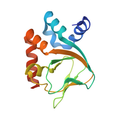
    MlotiK1 is a prokaryotic homolog of cyclic-nucleotide-dependent ion channels that contains an intracellular C-terminal cyclic nucleotide binding (CNB) domain. X-ray structures of the CNB domain have been solved in the absence of ligand and bound to cAMP. Both the full-length channel and CNB domain fragment are easily expressed and purified, making MlotiK1 a useful model system for dissecting activation by ligand binding. We have used X-ray crystallography to determine three new MlotiK1 CNB domain structures: a second apo configuration, a cGMP-bound structure, and a second cAMP-bound structure. In combination, the five MlotiK1 CNB domain structures provide a unique opportunity for analyzing, within a single protein, the structural differences between the apo state and the bound state, and the structural variability within each state. With this analysis as a guide, we have probed the nucleotide selectivity and importance of specific residue side chains in ligand binding and channel activation. These data help to identify ligand-protein interactions that are important for ligand dependence in MlotiK1 and, more globally, in the class of nucleotide-dependent proteins.


  • Organizational Affiliation

    Department of Molecular Biophysics and Biochemistry, Yale University, New Haven, CT 06520-8114, USA.


Macromolecules
Find similar proteins by:  (by identity cutoff)  |  3D Structure
Entity ID: 1
MoleculeChains Sequence LengthOrganismDetailsImage
Mll3241 proteinA,
B [auth C]
140N/AMutation(s): 1 
UniProt
Find proteins for Q98GN8 (Mesorhizobium japonicum (strain LMG 29417 / CECT 9101 / MAFF 303099))
Explore Q98GN8 
Go to UniProtKB:  Q98GN8
Entity Groups  
Sequence Clusters30% Identity50% Identity70% Identity90% Identity95% Identity100% Identity
UniProt GroupQ98GN8
Sequence Annotations
Expand
  • Reference Sequence
Experimental Data & Validation

Experimental Data

  • Method: X-RAY DIFFRACTION
  • Resolution: 2.00 Å
  • R-Value Free:  0.229 (Depositor), 0.220 (DCC) 
  • R-Value Work:  0.212 (Depositor), 0.210 (DCC) 
  • R-Value Observed: 0.213 (Depositor) 
Space Group: P 1 21 1
Unit Cell:
Length ( Å )Angle ( ˚ )
a = 34.535α = 90
b = 82.011β = 103.64
c = 50.119γ = 90
Software Package:
Software NamePurpose
REFMACrefinement
SCALEPACKdata scaling
CNSrefinement
PDB_EXTRACTdata extraction
DENZOdata reduction

Structure Validation

View Full Validation Report


Ligand Structure Quality Assessment 

Created with Raphaël 2.3.0Worse 01 BetterLigand structure goodness of fit to experimental dataBest fitted CMPClick on this verticalbar to view details

Entry History 

Deposition Data

Revision History  (Full details and data files)

  • Version 1.0: 2008-08-05
    Type: Initial release
  • Version 1.1: 2011-07-13
    Changes: Version format compliance
  • Version 1.2: 2017-10-25
    Changes: Refinement description
  • Version 1.3: 2019-07-24
    Changes: Data collection, Refinement description
  • Version 1.4: 2021-10-20
    Changes: Database references, Derived calculations
  • Version 1.5: 2024-02-21
    Changes: Data collection