3C7E | pdb_00003c7e

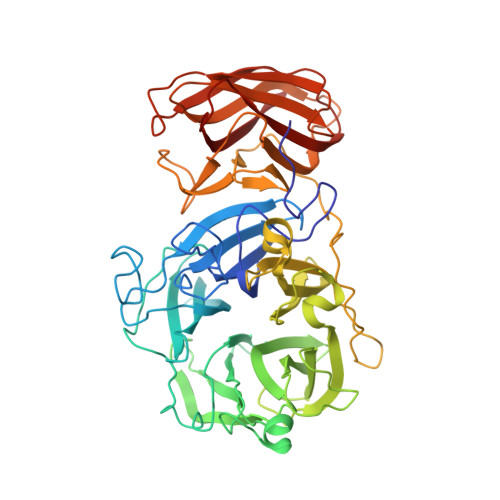
Crystal structure of a glycoside hydrolase family 43 arabinoxylan arabinofuranohydrolase from Bacillus subtilis.


Experimental Data Snapshot

  • Method: X-RAY DIFFRACTION
  • Resolution: 2.00 Å
  • R-Value Free: 
    0.213 (Depositor), 0.215 (DCC) 
  • R-Value Work: 
    0.168 (Depositor), 0.170 (DCC) 
  • R-Value Observed: 
    0.170 (Depositor) 

Starting Models: experimental
View more details

wwPDB Validation 3D Report Full Report

Validation slider image for 3C7E

This is version 1.3 of the entry. See complete history. 

Literature

Structural analysis of a glycoside hydrolase family 43 arabinoxylan arabinofuranohydrolase in complex with xylotetraose reveals a different binding mechanism compared with other members of the same family.

Vandermarliere, E., Bourgois, T.M., Winn, M.D., van Campenhout, S., Volckaert, G., Delcour, J.A., Strelkov, S.V., Rabijns, A., Courtin, C.M.

(2009) Biochem J 418: 39-47

  • DOI: https://doi.org/10.1042/BJ20081256
  • Primary Citation Related Structures: 
    3C7E, 3C7F, 3C7G, 3C7H, 3C7O

  • PubMed Abstract: 

    AXHs (arabinoxylan arabinofuranohydrolases) are alpha-L-arabinofuranosidases that specifically hydrolyse the glycosidic bond between arabinofuranosyl substituents and xylopyranosyl backbone residues of arabinoxylan. Bacillus subtilis was recently shown to produce an AXH that cleaves arabinose units from O-2- or O-3-mono-substituted xylose residues: BsAXH-m2,3 (B. subtilis AXH-m2,3). Crystallographic analysis reveals a two-domain structure for this enzyme: a catalytic domain displaying a five-bladed beta-propeller fold characteristic of GH (glycoside hydrolase) family 43 and a CBM (carbohydrate-binding module) with a beta-sandwich fold belonging to CBM family 6. Binding of substrate to BsAXH-m2,3 is largely based on hydrophobic stacking interactions, which probably allow the positional flexibility needed to hydrolyse both arabinose substituents at the O-2 or O-3 position of the xylose unit. Superposition of the BsAXH-m2,3 structure with known structures of the GH family 43 exo-acting enzymes, beta-xylosidase and alpha-L-arabinanase, each in complex with their substrate, reveals a different orientation of the sugar backbone.


  • Organizational Affiliation: 
    • Laboratory for Biocrystallography, Department of Pharmaceutical Sciences, Katholieke Universiteit Leuven, Herestraat 49, O&N II, bus 822, 3000 Leuven, Belgium.

Macromolecule Content 

  • Total Structure Weight: 51.95 kDa 
  • Atom Count: 3,997 
  • Modeled Residue Count: 487 
  • Deposited Residue Count: 487 
  • Unique protein chains: 1

Macromolecules

Find similar proteins by:|  3D Structure
Entity ID: 1
MoleculeChains  Sequence LengthOrganismDetailsImage
Endo-1,4-beta-xylanase487Bacillus subtilisMutation(s): 0 
Gene Names: xynD
EC: 3.2.1.55
UniProt
Find proteins for Q45071 (Bacillus subtilis (strain 168))
Explore Q45071 
Go to UniProtKB:  Q45071
Entity Groups
Sequence Clusters30% Identity50% Identity70% Identity90% Identity95% Identity100% Identity
UniProt GroupQ45071
Sequence Annotations
Expand
Reference Sequence

Small Molecules

Ligands 3 Unique
IDChains Name / Formula / InChI Key2D Diagram3D Interactions
FMT

Query on FMT


Download:Ideal Coordinates CCD File
E [auth A],
F [auth A],
G [auth A],
H [auth A]
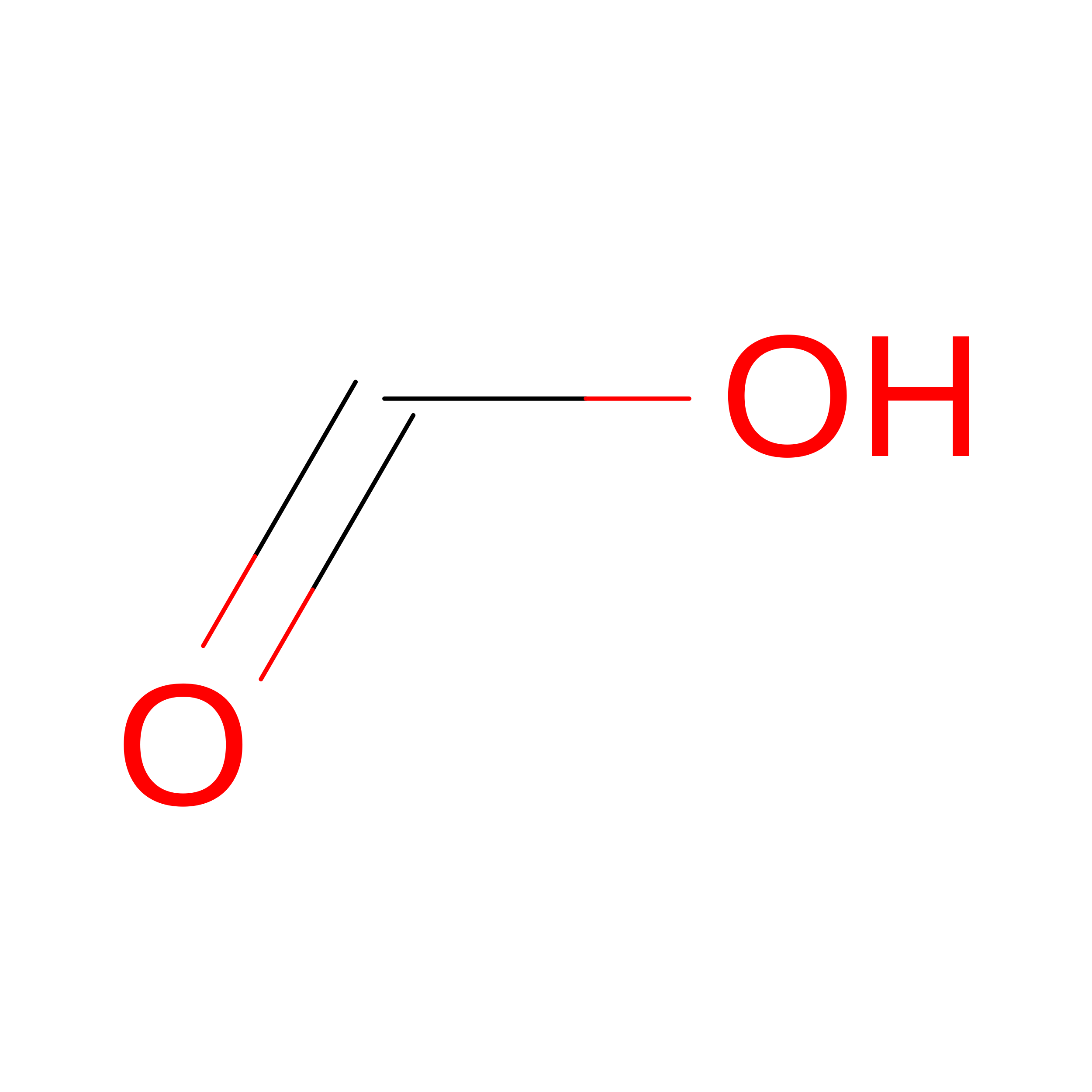
FORMIC ACID
C H2 O2
BDAGIHXWWSANSR-UHFFFAOYSA-N
CA

Query on CA


Download:Ideal Coordinates CCD File
B [auth A]CALCIUM ION
Ca
BHPQYMZQTOCNFJ-UHFFFAOYSA-N
NA

Query on NA


Download:Ideal Coordinates CCD File
C [auth A],
D [auth A]
SODIUM ION
Na
FKNQFGJONOIPTF-UHFFFAOYSA-N

Experimental Data & Validation

Experimental Data

  • Method: X-RAY DIFFRACTION
  • Resolution: 2.00 Å
  • R-Value Free:  0.213 (Depositor), 0.215 (DCC) 
  • R-Value Work:  0.168 (Depositor), 0.170 (DCC) 
  • R-Value Observed: 0.170 (Depositor) 
Space Group: P 21 21 21
Unit Cell:
Length ( Å )Angle ( ˚ )
a = 67.71α = 90
b = 74.43β = 90
c = 106.99γ = 90
Software Package:
Software NamePurpose
REFMACrefinement
MOSFLMdata reduction
SCALAdata scaling
PHASERphasing

Structure Validation

View Full Validation Report


Entry History 

Revision History  (Full details and data files)

  • Version 1.0: 2008-11-18
    Type: Initial release
  • Version 1.1: 2011-07-13
    Changes: Version format compliance
  • Version 1.2: 2017-08-23
    Changes: Data collection
  • Version 1.3: 2023-08-30
    Changes: Data collection, Database references, Derived calculations, Refinement description