3C6B | pdb_00003c6b

Reaction product of paraoxon and S-formylglutathione hydrolase W197I mutant


Experimental Data Snapshot

  • Method: X-RAY DIFFRACTION
  • Resolution: 2.17 Å
  • R-Value Free: 
    0.252 (Depositor), 0.249 (DCC) 
  • R-Value Work: 
    0.190 (Depositor), 0.191 (DCC) 
  • R-Value Observed: 
    0.193 (Depositor) 

Starting Model: experimental
View more details

wwPDB Validation 3D Report Full Report

Validation slider image for 3C6B

This is version 1.5 of the entry. See complete history

Literature

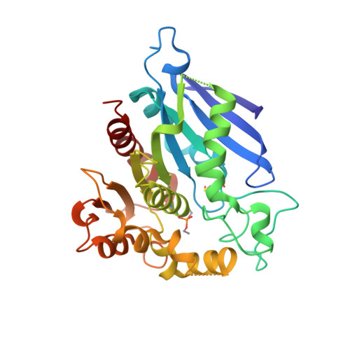
Structural characterization and reversal of the natural organophosphate resistance of a D-type esterase, Saccharomyces cerevisiae S-formylglutathione hydrolase.

Legler, P.M.Kumaran, D.Swaminathan, S.Studier, F.W.Millard, C.B.

(2008) Biochemistry 47: 9592-9601

  • DOI: https://doi.org/10.1021/bi8010016
  • Primary Citation Related Structures: 
    1PV1, 3C6B

  • PubMed Abstract: 

    Saccharomyces cerevisiae expresses a 67.8 kDa homodimeric serine thioesterase, S-formylglutathione hydrolase (SFGH), that is 39.9% identical with human esterase D. Both enzymes possess significant carboxylesterase and S-formylglutathione thioesterase activity but are unusually resistant to organophosphate (OP) inhibitors. We determined the X-ray crystal structure of yeast (y) SFGH to 2.3 A resolution by multiwavelength anomalous dispersion and used the structure to guide site-specific mutagenesis experiments addressing substrate and inhibitor reactivity. Our results demonstrate a steric mechanism of OP resistance mediated by a single indole ring (W197) located in an enzyme "acyl pocket". The W197I substitution enhances ySFGH reactivity with paraoxon by >1000-fold ( k i (W197I) = 16 +/- 2 mM (-1) h (-1)), thereby overcoming natural OP resistance. W197I increases the rate of OP inhibition under pseudo-first-order conditions but does not accelerate OP hydrolysis. The structure of the paraoxon-inhibited W197I variant was determined by molecular replacement (2.2 A); it revealed a stabilized sulfenic acid at Cys60. Wild-type (WT) ySFGH is inhibited by thiol reactive compounds and is sensitive to oxidation; thus, the cysteine sulfenic acid may play a role in the regulation of a "D-type" esterase. The structure of the W197I variant is the first reported cysteine sulfenic acid in a serine esterase. We constructed five Cys60/W197I variants and show that introducing a positive charge near the oxyanion hole, W197I/C60R or W197I/C60K, results in a further enhancement of the rates of phosphorylation with paraoxon ( k i = 42 or 80 mM (-1) h (-1), respectively) but does not affect the dephosphorylation of the enzyme. We also characterized three histidine substitutions near the oxyanion hole, G57H, L58H, and M162H, which significantly decrease esterase activity.


  • Organizational Affiliation
    • Division of Biochemistry, Walter Reed Army Institute of Research, Silver Spring, Maryland 20910, USA.

Macromolecule Content 

  • Total Structure Weight: 34.06 kDa 
  • Atom Count: 2,382 
  • Modeled Residue Count: 291 
  • Deposited Residue Count: 299 
  • Unique protein chains: 1

Macromolecules

Find similar proteins by:|  3D Structure
Entity ID: 1
MoleculeChains  Sequence LengthOrganismDetailsImage
S-formylglutathione hydrolase299Saccharomyces cerevisiaeMutation(s): 1 
Gene Names: YJL068c
EC: 3.1.2.12
UniProt
Find proteins for P40363 (Saccharomyces cerevisiae (strain ATCC 204508 / S288c))
Explore P40363 
Go to UniProtKB:  P40363
Entity Groups
Sequence Clusters30% Identity50% Identity70% Identity90% Identity95% Identity100% Identity
UniProt GroupP40363
Sequence Annotations
Expand
Reference Sequence

Small Molecules

Modified Residues  2 Unique
IDChains TypeFormula2D DiagramParent
CSO
Query on CSO
A
L-PEPTIDE LINKINGC3 H7 N O3 SCYS
SDP
Query on SDP
A
L-PEPTIDE LINKINGC7 H16 N O6 PSER

Experimental Data & Validation

Experimental Data

  • Method: X-RAY DIFFRACTION
  • Resolution: 2.17 Å
  • R-Value Free:  0.252 (Depositor), 0.249 (DCC) 
  • R-Value Work:  0.190 (Depositor), 0.191 (DCC) 
  • R-Value Observed: 0.193 (Depositor) 
Space Group: C 1 2 1
Unit Cell:
Length ( Å )Angle ( ˚ )
a = 57.902α = 90
b = 68.99β = 92.58
c = 78.993γ = 90
Software Package:
Software NamePurpose
REFMACrefinement
PROTEUM PLUSdata collection
SAINTdata reduction
PROTEUM PLUSdata scaling
AMoREphasing

Structure Validation

View Full Validation Report


Entry History 

Deposition Data

Revision History  (Full details and data files)

  • Version 1.0: 2008-08-12
    Type: Initial release
  • Version 1.1: 2011-07-13
    Changes: Version format compliance
  • Version 1.2: 2018-04-04
    Changes: Data collection, Source and taxonomy
  • Version 1.3: 2021-10-20
    Changes: Database references, Derived calculations
  • Version 1.4: 2023-08-30
    Changes: Data collection, Refinement description
  • Version 1.5: 2023-11-15
    Changes: Data collection