3BYQ | pdb_00003byq

Crystal structure of a duf1185 family protein (bb2672) from bordetella bronchiseptica rb50 at 1.70 A resolution


Experimental Data Snapshot

  • Method: X-RAY DIFFRACTION
  • Resolution: 1.70 Å
  • R-Value Free: 
    0.169 (Depositor), 0.165 (DCC) 
  • R-Value Work: 
    0.138 (Depositor), 0.139 (DCC) 
  • R-Value Observed: 
    0.139 (Depositor) 

wwPDB Validation 3D Report Full Report

Validation slider image for 3BYQ

This is version 1.5 of the entry. See complete history

Literature

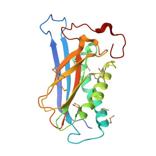
Structures of the first representatives of Pfam family PF06684 (DUF1185) reveal a novel variant of the Bacillus chorismate mutase fold and suggest a role in amino-acid metabolism.

Bakolitsa, C.Kumar, A.Jin, K.K.McMullan, D.Krishna, S.S.Miller, M.D.Abdubek, P.Acosta, C.Astakhova, T.Axelrod, H.L.Burra, P.Carlton, D.Chen, C.Chiu, H.J.Clayton, T.Das, D.Deller, M.C.Duan, L.Elias, Y.Ellrott, K.Ernst, D.Farr, C.L.Feuerhelm, J.Grant, J.C.Grzechnik, A.Grzechnik, S.K.Han, G.W.Jaroszewski, L.Johnson, H.A.Klock, H.E.Knuth, M.W.Kozbial, P.Marciano, D.Morse, A.T.Murphy, K.D.Nigoghossian, E.Nopakun, A.Okach, L.Paulsen, J.Puckett, C.Reyes, R.Rife, C.L.Sefcovic, N.Tien, H.J.Trame, C.B.Trout, C.V.van den Bedem, H.Weekes, D.White, A.Xu, Q.Hodgson, K.O.Wooley, J.Elsliger, M.A.Deacon, A.M.Godzik, A.Lesley, S.A.Wilson, I.A.

(2010) Acta Crystallogr Sect F Struct Biol Cryst Commun 66: 1182-1189

  • DOI: https://doi.org/10.1107/S1744309109050647
  • Primary Citation Related Structures: 
    2QTP, 3BYQ

  • PubMed Abstract: 

    The crystal structures of BB2672 and SPO0826 were determined to resolutions of 1.7 and 2.1 Å by single-wavelength anomalous dispersion and multiple-wavelength anomalous dispersion, respectively, using the semi-automated high-throughput pipeline of the Joint Center for Structural Genomics (JCSG) as part of the NIGMS Protein Structure Initiative (PSI). These proteins are the first structural representatives of the PF06684 (DUF1185) Pfam family. Structural analysis revealed that both structures adopt a variant of the Bacillus chorismate mutase fold (BCM). The biological unit of both proteins is a hexamer and analysis of homologs indicates that the oligomer interface residues are highly conserved. The conformation of the critical regions for oligomerization appears to be dependent on pH or salt concentration, suggesting that this protein might be subject to environmental regulation. Structural similarities to BCM and genome-context analysis suggest a function in amino-acid synthesis.


  • Organizational Affiliation
    • Stanford Synchrotron Radiation Lightsource, SLAC National Accelerator Laboratory, Menlo Park, CA, USA.

Macromolecule Content 

  • Total Structure Weight: 66.43 kDa 
  • Atom Count: 5,150 
  • Modeled Residue Count: 574 
  • Deposited Residue Count: 579 
  • Unique protein chains: 1

Macromolecules

Find similar proteins by:|  3D Structure
Entity ID: 1
MoleculeChains  Sequence LengthOrganismDetailsImage
Uncharacterized protein DUF1185
A, B, C
193Bordetella bronchiseptica RB50Mutation(s): 0 
Gene Names: NP_889209.1BB2672
UniProt
Find proteins for A0A0H3LTD0 (Bordetella bronchiseptica (strain ATCC BAA-588 / NCTC 13252 / RB50))
Explore A0A0H3LTD0 
Go to UniProtKB:  A0A0H3LTD0
Entity Groups
Sequence Clusters30% Identity50% Identity70% Identity90% Identity95% Identity100% Identity
UniProt GroupA0A0H3LTD0
Sequence Annotations
Expand
Reference Sequence

Small Molecules

Ligands 3 Unique
IDChains Name / Formula / InChI Key2D Diagram3D Interactions
PG4

Query on PG4


Download:Ideal Coordinates CCD File
BA [auth C],
E [auth A]
TETRAETHYLENE GLYCOL
C8 H18 O5
UWHCKJMYHZGTIT-UHFFFAOYSA-N
EDO

Query on EDO


Download:Ideal Coordinates CCD File
CA [auth C]
DA [auth C]
EA [auth C]
F [auth A]
FA [auth C]
CA [auth C],
DA [auth C],
EA [auth C],
F [auth A],
FA [auth C],
G [auth A],
GA [auth C],
H [auth A],
HA [auth C],
I [auth A],
IA [auth C],
J [auth A],
JA [auth C],
K [auth A],
L [auth A],
M [auth A],
O [auth B],
P [auth B],
Q [auth B],
R [auth B],
S [auth B],
T [auth B],
U [auth B],
V [auth B],
W [auth B],
X [auth B],
Y [auth B]
1,2-ETHANEDIOL
C2 H6 O2
LYCAIKOWRPUZTN-UHFFFAOYSA-N
CL

Query on CL


Download:Ideal Coordinates CCD File
AA [auth C],
D [auth A],
N [auth B],
Z [auth C]
CHLORIDE ION
Cl
VEXZGXHMUGYJMC-UHFFFAOYSA-M
Modified Residues  1 Unique
IDChains TypeFormula2D DiagramParent
MSE
Query on MSE
A, B, C
L-PEPTIDE LINKINGC5 H11 N O2 SeMET

Experimental Data & Validation

Experimental Data

  • Method: X-RAY DIFFRACTION
  • Resolution: 1.70 Å
  • R-Value Free:  0.169 (Depositor), 0.165 (DCC) 
  • R-Value Work:  0.138 (Depositor), 0.139 (DCC) 
  • R-Value Observed: 0.139 (Depositor) 
Space Group: C 2 2 21
Unit Cell:
Length ( Å )Angle ( ˚ )
a = 96.542α = 90
b = 133.127β = 90
c = 92.544γ = 90
Software Package:
Software NamePurpose
REFMACrefinement
PHENIXrefinement
SHELXphasing
MolProbitymodel building
SCALAdata scaling
PDB_EXTRACTdata extraction
MAR345data collection
MOSFLMdata reduction
SHELXDphasing
autoSHARPphasing

Structure Validation

View Full Validation Report


Entry History 

Deposition Data

Revision History  (Full details and data files)

  • Version 1.0: 2008-01-29
    Type: Initial release
  • Version 1.1: 2011-07-13
    Changes: Advisory, Version format compliance
  • Version 1.2: 2017-10-25
    Changes: Author supporting evidence, Refinement description
  • Version 1.3: 2019-07-24
    Changes: Data collection, Derived calculations, Refinement description
  • Version 1.4: 2023-02-01
    Changes: Database references, Derived calculations
  • Version 1.5: 2024-10-16
    Changes: Data collection, Structure summary