3BS1 | pdb_00003bs1

Structure of the Staphylococcus aureus AgrA LytTR Domain Bound to DNA Reveals a Beta Fold with a Novel Mode of Binding


Experimental Data Snapshot

  • Method: X-RAY DIFFRACTION
  • Resolution: 1.60 Å
  • R-Value Free: 
    0.221 (Depositor), 0.231 (DCC) 
  • R-Value Work: 
    0.195 (Depositor), 0.210 (DCC) 
  • R-Value Observed: 
    0.197 (Depositor) 

wwPDB Validation 3D Report Full Report

Validation slider image for 3BS1

This is version 1.2 of the entry. See complete history

Literature

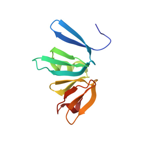
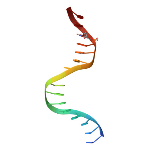
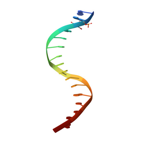
Structure of the Staphylococcus aureus AgrA LytTR Domain Bound to DNA Reveals a Beta Fold with an Unusual Mode of Binding.

Sidote, D.J.Barbieri, C.M.Wu, T.Stock, A.M.

(2008) Structure 16: 727-735

  • DOI: https://doi.org/10.1016/j.str.2008.02.011
  • Primary Citation Related Structures: 
    3BS1

  • PubMed Abstract: 

    The LytTR domain is a DNA-binding motif found within the AlgR/AgrA/LytR family of transcription factors that regulate virulence factor and toxin gene expression in pathogenic bacteria. This previously uncharacterized domain lacks sequence similarity with proteins of known structure. The crystal structure of the DNA-binding domain of Staphylococcus aureus AgrA complexed with a DNA pentadecamer duplex has been determined at 1.6 A resolution. The structure establishes a 10-stranded beta fold for the LytTR domain and reveals its mode of interaction with DNA. Residues within loop regions of AgrA contact two successive major grooves and the intervening minor groove on one face of the oligonucleotide duplex, inducing a substantial bend in the DNA. Loss of DNA binding upon substitution of key interacting residues in AgrA supports the observed binding mode. This mode of protein-DNA interaction provides a potential target for future antimicrobial drug design.


  • Organizational Affiliation
    • Center for Advanced Biotechnology and Medicine and Department of Biochemistry, Howard Hughes Medical Institute, University of Medicine and Dentistry of New Jersey, Robert Wood Johnson Medical School, 679 Hoes Lane, Piscataway, NJ 08854, USA.

Macromolecule Content 

  • Total Structure Weight: 22.33 kDa 
  • Atom Count: 1,747 
  • Modeled Residue Count: 135 
  • Deposited Residue Count: 135 
  • Unique protein chains: 1
  • Unique nucleic acid chains: 2

Macromolecules


Find similar proteins by:|  3D Structure
Entity ID: 3
MoleculeChains  Sequence LengthOrganismDetailsImage
Accessory gene regulator protein AC [auth A]103Staphylococcus aureusMutation(s): 0 
Gene Names: agrAagr
UniProt
Find proteins for P0A0I7 (Staphylococcus aureus)
Explore P0A0I7 
Go to UniProtKB:  P0A0I7
Entity Groups
Sequence Clusters30% Identity50% Identity70% Identity90% Identity95% Identity100% Identity
UniProt GroupP0A0I7
Sequence Annotations
Expand
Reference Sequence
Find similar nucleic acids by:  Sequence
Entity ID: 1
MoleculeChains LengthOrganismImage
DNA (5'-D(*DTP*DTP*DTP*DAP*DAP*DCP*DAP*DGP*DTP*DTP*DAP*DAP*DGP*(BRU)P*DAP*DT)-3')A [auth B]16N/A
Sequence Annotations
Expand
Reference Sequence
Find similar nucleic acids by:  Sequence
Entity ID: 2
MoleculeChains LengthOrganismImage
DNA (5'-D(*DAP*DAP*(BRU)P*DAP*DCP*DTP*DTP*DAP*DAP*DCP*DTP*DGP*DTP*DTP*DAP*DA)-3')B [auth C]16N/A
Sequence Annotations
Expand
Reference Sequence

Experimental Data & Validation

Experimental Data

  • Method: X-RAY DIFFRACTION
  • Resolution: 1.60 Å
  • R-Value Free:  0.221 (Depositor), 0.231 (DCC) 
  • R-Value Work:  0.195 (Depositor), 0.210 (DCC) 
  • R-Value Observed: 0.197 (Depositor) 
Space Group: P 41
Unit Cell:
Length ( Å )Angle ( ˚ )
a = 47.938α = 90
b = 47.938β = 90
c = 100.112γ = 90
Software Package:
Software NamePurpose
REFMACrefinement
ADSCdata collection
HKL-2000data reduction
HKL-2000data scaling
SHARPphasing

Structure Validation

View Full Validation Report


Entry History 

Deposition Data

Revision History  (Full details and data files)

  • Version 1.0: 2008-04-08
    Type: Initial release
  • Version 1.1: 2011-07-13
    Changes: Version format compliance
  • Version 1.2: 2024-02-21
    Changes: Data collection, Database references, Derived calculations