3BMP | pdb_00003bmp

HUMAN BONE MORPHOGENETIC PROTEIN-2 (BMP-2)


Experimental Data Snapshot

  • Method: X-RAY DIFFRACTION
  • Resolution: 2.70 Å
  • R-Value Free: 
    0.278 (Depositor) 
  • R-Value Work: 
    0.242 (Depositor), 0.255 (DCC) 

Starting Model: experimental
View more details

wwPDB Validation 3D Report Full Report

Validation slider image for 3BMP

This is version 1.5 of the entry. See complete history

Literature

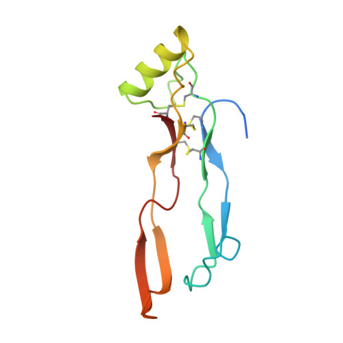
Crystal structure of human bone morphogenetic protein-2 at 2.7 A resolution.

Scheufler, C.Sebald, W.Hulsmeyer, M.

(1999) J Mol Biology 287: 103-115

  • DOI: https://doi.org/10.1006/jmbi.1999.2590
  • Primary Citation Related Structures: 
    3BMP

  • PubMed Abstract: 

    Homodimeric bone morphogenetic protein-2 (BMP-2) is a member of the transforming growth factor beta (TGF-beta) superfamily that induces bone formation and regeneration, and determines important steps during early stages of embryonic development in vertebrates and non-vertebrates. BMP-2 can interact with two types of receptor chains, as well as with proteins of the extracellular matrix and several regulatory proteins. We report here the crystal structure of human BMP-2 determined by molecular replacement and refined to an R-value of 24.2 % at 2.7 A resolution. A common scaffold of BMP-2, BMP-7 and the TGF-betas, i.e. the cystine-knot motif and two finger-like double-stranded beta-sheets, can be superimposed with r. m.s. deviations of around 1 A. In contrast to the TGF-betas, the structure of BMP-2 shows differences in the flexibility of the N terminus and the orientation of the central alpha-helix as well as two external loops at the fingertips with respect to the scaffold. This is also known from the BMP-7 model. Small secondary structure elements in the loop regions of BMP-2 and BMP-7 seem to be specific for the respective BMP-subgroup. Two identical helix-finger clefts and two distinct cavities located around the central 2-fold axis of the dimer show characteristic shapes, polarity and surface charges. The possible function of these specific features in the interaction of BMP-2 with its binding partners is discussed.


  • Organizational Affiliation
    • Physiological Chemistry II, University of Würzburg, Würzburg, 97074, Germany.

Macromolecule Content 

  • Total Structure Weight: 13.04 kDa 
  • Atom Count: 879 
  • Modeled Residue Count: 106 
  • Deposited Residue Count: 114 
  • Unique protein chains: 1

Macromolecules

Find similar proteins by:|  3D Structure
Entity ID: 1
MoleculeChains  Sequence LengthOrganismDetailsImage
PROTEIN (BONE MORPHOGENETIC PROTEIN 2 (BMP-2))114Homo sapiensMutation(s): 0 
UniProt & NIH Common Fund Data Resources
Find proteins for P12643 (Homo sapiens)
Explore P12643 
Go to UniProtKB:  P12643
PHAROS:  P12643
GTEx:  ENSG00000125845 
Entity Groups
Sequence Clusters30% Identity50% Identity70% Identity90% Identity95% Identity100% Identity
UniProt GroupP12643
Sequence Annotations
Expand
Reference Sequence

Small Molecules

Ligands 1 Unique
IDChains Name / Formula / InChI Key2D Diagram3D Interactions
MPD

Query on MPD


Download:Ideal Coordinates CCD File
B [auth A](4S)-2-METHYL-2,4-PENTANEDIOL
C6 H14 O2
SVTBMSDMJJWYQN-YFKPBYRVSA-N

Experimental Data & Validation

Experimental Data

  • Method: X-RAY DIFFRACTION
  • Resolution: 2.70 Å
  • R-Value Free:  0.278 (Depositor) 
  • R-Value Work:  0.242 (Depositor), 0.255 (DCC) 
Space Group: H 3 2
Unit Cell:
Length ( Å )Angle ( ˚ )
a = 91.44α = 90
b = 91.44β = 90
c = 107.75γ = 120
Software Package:
Software NamePurpose
XDSdata scaling
XDSdata reduction
AMoREphasing
CNSrefinement

Structure Validation

View Full Validation Report


Entry History 

Deposition Data

Revision History  (Full details and data files)

  • Version 1.0: 2000-03-12
    Type: Initial release
  • Version 1.1: 2008-04-26
    Changes: Version format compliance
  • Version 1.2: 2011-07-13
    Changes: Derived calculations, Version format compliance
  • Version 1.3: 2018-04-04
    Changes: Data collection
  • Version 1.4: 2023-08-30
    Changes: Data collection, Database references, Derived calculations, Refinement description
  • Version 1.5: 2024-11-13
    Changes: Structure summary