3BCD | pdb_00003bcd

Alpha-amylase B in complex with maltotetraose and alpha-cyclodextrin


Experimental Data Snapshot

  • Method: X-RAY DIFFRACTION
  • Resolution: 2.20 Å
  • R-Value Free: 
    0.240 (Depositor), 0.251 (DCC) 
  • R-Value Work: 
    0.196 (Depositor), 0.218 (DCC) 
  • R-Value Observed: 
    0.197 (Depositor) 

Starting Model: experimental
View more details

wwPDB Validation 3D Report Full Report

Validation slider image for 3BCD

This is version 2.1 of the entry. See complete history

Literature

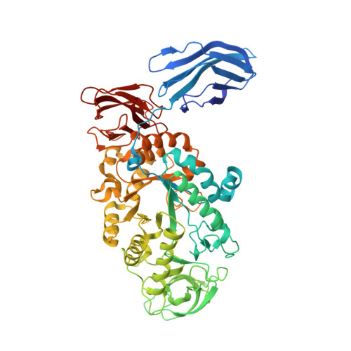
Crystal Structure of the Polyextremophilic alpha-Amylase AmyB from Halothermothrix orenii: Details of a Productive Enzyme-Substrate Complex and an N Domain with a Role in Binding Raw Starch

Tan, T.-C.Mijts, B.N.Swaminathan, K.Patel, B.K.C.Divne, C.

(2008) J Mol Biology 378: 850-868

  • DOI: https://doi.org/10.1016/j.jmb.2008.02.041
  • Primary Citation Related Structures: 
    3BC9, 3BCD, 3BCF

  • PubMed Abstract: 

    The gene for a membrane-bound, halophilic, and thermostable alpha-amylase, AmyB, from Halothermothrix orenii was cloned and sequenced. The crystal structure shows that, in addition to the typical domain organization of family 13 glycoside hydrolases, AmyB carries an additional N-terminal domain (N domain) that forms a large groove--the N-C groove--some 30 A away from the active site. The structure of AmyB with the inhibitor acarbose at 1.35 A resolution shows that a nonasaccharide has been synthesized through successive transglycosylation reactions of acarbose. Unexpectedly, in a complex of wild-type AmyB with alpha-cyclodextrin and maltoheptaose at 2.2 A resolution, a maltotetraose molecule is bound in subsites -1 to +3, spanning the cleavage point at -1/+1, with the -1 glucosyl residue present as a (2)S(o) skew boat. This wild-type AmyB complex was obtained in the presence of a large excess of substrate, a condition under which it is possible to capture Michaelis complexes, which may explain the observed binding across -1/+1 and ring distortion. We observe three methionine side chains that serve as "binding platforms" for glucosyl rings in AmyB, a seemingly rare occurrence in carbohydrate-binding proteins. The structures and results from the biochemical characterization of AmyB and AmyB lacking the N domain show that the N domain increases binding of the enzyme to raw starch. Furthermore, theoretical modeling suggests that the N-C groove can accommodate, spatially and chemically, large substrates such as A-starch.


  • Organizational Affiliation
    • KTH School of Biotechnology, AlbaNova University Center, Roslagstullsbacken 21, SE-106 91 Stockholm, Sweden.

Macromolecule Content 

  • Total Structure Weight: 71.21 kDa 
  • Atom Count: 5,170 
  • Modeled Residue Count: 585 
  • Deposited Residue Count: 599 
  • Unique protein chains: 1

Macromolecules

Find similar proteins by:|  3D Structure
Entity ID: 1
MoleculeChains  Sequence LengthOrganismDetailsImage
Alpha amylase, catalytic region599Halothermothrix orenii H 168Mutation(s): 0 
Gene Names: amyb
EC: 3.2.1.1 (PDB Primary Data), 3.2.1.98 (UniProt)
UniProt
Find proteins for B8CZ54 (Halothermothrix orenii (strain H 168 / OCM 544 / DSM 9562))
Explore B8CZ54 
Go to UniProtKB:  B8CZ54
Entity Groups
Sequence Clusters30% Identity50% Identity70% Identity90% Identity95% Identity100% Identity
UniProt GroupB8CZ54
Sequence Annotations
Expand
Reference Sequence

Oligosaccharides

Help  
Entity ID: 2
MoleculeChains Length2D Diagram GlycosylationD Interactions
alpha-D-glucopyranose-(1-4)-alpha-D-glucopyranose-(1-4)-alpha-D-glucopyranose-(1-4)-beta-D-glucopyranose
B
4N/A
Glycosylation Resources
GlyTouCan: G69211UR
GlyCosmos: G69211UR
GlyGen: G69211UR
Entity ID: 3
MoleculeChains Length2D Diagram GlycosylationD Interactions
alpha-D-glucopyranose-(1-4)-alpha-D-glucopyranose-(1-4)-alpha-D-glucopyranose-(1-4)-alpha-D-glucopyranose
C
4N/A
Glycosylation Resources
GlyTouCan: G87171PZ
GlyCosmos: G87171PZ
GlyGen: G87171PZ
Entity ID: 4
MoleculeChains Length2D Diagram GlycosylationD Interactions
Cyclohexakis-(1-4)-(alpha-D-glucopyranose)
D
6N/A
Glycosylation Resources
GlyTouCan: G22307HL
GlyCosmos: G22307HL

Small Molecules

Ligands 2 Unique
IDChains Name / Formula / InChI Key2D Diagram3D Interactions
CA

Query on CA


Download:Ideal Coordinates CCD File
E [auth A]
F [auth A]
G [auth A]
I [auth A]
J [auth A]
E [auth A],
F [auth A],
G [auth A],
I [auth A],
J [auth A],
K [auth A],
L [auth A]
CALCIUM ION
Ca
BHPQYMZQTOCNFJ-UHFFFAOYSA-N
NA

Query on NA


Download:Ideal Coordinates CCD File
H [auth A]SODIUM ION
Na
FKNQFGJONOIPTF-UHFFFAOYSA-N

Biologically Interesting Molecules (External Reference) 

2 Unique

Experimental Data & Validation

Experimental Data

  • Method: X-RAY DIFFRACTION
  • Resolution: 2.20 Å
  • R-Value Free:  0.240 (Depositor), 0.251 (DCC) 
  • R-Value Work:  0.196 (Depositor), 0.218 (DCC) 
  • R-Value Observed: 0.197 (Depositor) 
Space Group: C 1 2 1
Unit Cell:
Length ( Å )Angle ( ˚ )
a = 228.7α = 90
b = 78.05β = 98.9
c = 50.64γ = 90
Software Package:
Software NamePurpose
REFMACrefinement
MAR345data collection
XDSdata reduction
XSCALEdata scaling
REFMACphasing

Structure Validation

View Full Validation Report


Entry History 

Deposition Data

Revision History  (Full details and data files)

  • Version 1.0: 2008-04-22
    Type: Initial release
  • Version 1.1: 2011-07-13
    Changes: Advisory, Source and taxonomy, Version format compliance
  • Version 1.2: 2017-10-25
    Changes: Data collection, Refinement description
  • Version 2.0: 2020-07-29
    Type: Remediation
    Reason: Carbohydrate remediation
    Changes: Atomic model, Data collection, Derived calculations, Non-polymer description, Structure summary
  • Version 2.1: 2023-11-01
    Changes: Data collection, Database references, Refinement description, Structure summary