3B1D | pdb_00003b1d

Crystal structure of betaC-S lyase from Streptococcus anginosus in complex with L-serine: External aldimine form


Experimental Data Snapshot

  • Method: X-RAY DIFFRACTION
  • Resolution: 1.66 Å
  • R-Value Free: 
    0.177 (Depositor), 0.183 (DCC) 
  • R-Value Work: 
    0.151 (Depositor), 0.160 (DCC) 
  • R-Value Observed: 
    0.152 (Depositor) 

wwPDB Validation 3D Report Full Report

Validation slider image for 3B1D

Ligand Structure Quality Assessment 


This is version 1.3 of the entry. See complete history

Literature

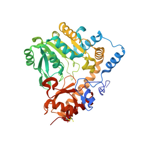
Structural insights into catalysis by beta C-S lyase from Streptococcus anginosus

Kezuka, Y.Yoshida, Y.Nonaka, T.

(2012) Proteins 80: 2447-2458

  • DOI: https://doi.org/10.1002/prot.24129
  • Primary Citation Related Structures: 
    3B1C, 3B1D, 3B1E

  • PubMed Abstract: 

    Hydrogen sulfide (H(2)S) is a causative agent of oral malodor and may play an important role in the pathogenicity of oral bacteria such as Streptococcus anginosus. In this microorganism, H(2)S production is associated with βC-S lyase (Lcd) encoded by lcd gene, which is a pyridoxal 5'-phosphate (PLP)-dependent enzyme that catalyzes the α,β-elimination of sulfur-containing amino acids. When Lcd acts on L-cysteine, H(2)S is produced along with pyruvate and ammonia. To understand the H(2)S-producing mechanism of Lcd in detail, we determined the crystal structures of substrate-free Lcd (internal aldimine form) and two reaction intermediate complexes (external aldimine and α-aminoacrylate forms). The formation of intermediates induced little changes in the overall structure of the enzyme and in the active site residues, with the exception of Lys234, a PLP-binding residue. Structural and mutational analyses highlighted the importance of the active site residues Tyr60, Tyr119, and Arg365. In particular, Tyr119 forms a hydrogen bond with the side chain oxygen atom of L-serine, a substrate analog, in the external aldimine form suggesting its role in the recognition of the sulfur atom of the true substrate (L-cysteine). Tyr119 also plays a role in fixing the PLP cofactor at the proper position during catalysis through binding with its side chain. Finally, we partly modified the catalytic mechanism known for cystalysin, a βC-S lyase from Treponema denticola, and proposed an improved mechanism, which seems to be common to the βC-S lyases from oral bacteria.


  • Organizational Affiliation
    • Department of Structural Biology, School of Pharmacy, Iwate Medical University, Yahaba, Iwate, Japan.

Macromolecule Content 

  • Total Structure Weight: 183.15 kDa 
  • Atom Count: 14,658 
  • Modeled Residue Count: 1,546 
  • Deposited Residue Count: 1,568 
  • Unique protein chains: 1

Macromolecules

Find similar proteins by:|  3D Structure
Entity ID: 1
MoleculeChains  Sequence LengthOrganismDetailsImage
BetaC-S lyase
A, B, C, D
392Streptococcus anginosusMutation(s): 0 
Gene Names: lcd
EC: 4.4.1.8 (PDB Primary Data), 4.4.1.13 (UniProt)
UniProt
Find proteins for A6BMJ3 (Streptococcus anginosus)
Explore A6BMJ3 
Go to UniProtKB:  A6BMJ3
Entity Groups
Sequence Clusters30% Identity50% Identity70% Identity90% Identity95% Identity100% Identity
UniProt GroupA6BMJ3
Sequence Annotations
Expand
Reference Sequence

Small Molecules

Ligands 4 Unique
IDChains Name / Formula / InChI Key2D Diagram3D Interactions
PLS

Query on PLS


Download:Ideal Coordinates CCD File
F [auth A],
K [auth B],
P [auth C],
U [auth D]
[3-HYDROXY-2-METHYL-5-PHOSPHONOOXYMETHYL-PYRIDIN-4-YLMETHYL]-SERINE
C11 H17 N2 O8 P
ODVKKQWXKRZJLG-VIFPVBQESA-N
PLP

Query on PLP


Download:Ideal Coordinates CCD File
E [auth A],
O [auth C]
PYRIDOXAL-5'-PHOSPHATE
C8 H10 N O6 P
NGVDGCNFYWLIFO-UHFFFAOYSA-N
EPE

Query on EPE


Download:Ideal Coordinates CCD File
G [auth A]
H [auth A]
I [auth A]
L [auth B]
M [auth B]
G [auth A],
H [auth A],
I [auth A],
L [auth B],
M [auth B],
Q [auth C],
R [auth C],
S [auth C],
V [auth D],
W [auth D],
X [auth D]
4-(2-HYDROXYETHYL)-1-PIPERAZINE ETHANESULFONIC ACID
C8 H18 N2 O4 S
JKMHFZQWWAIEOD-UHFFFAOYSA-N
NA

Query on NA


Download:Ideal Coordinates CCD File
J [auth A],
N [auth B],
T [auth C],
Y [auth D]
SODIUM ION
Na
FKNQFGJONOIPTF-UHFFFAOYSA-N

Experimental Data & Validation

Experimental Data

  • Method: X-RAY DIFFRACTION
  • Resolution: 1.66 Å
  • R-Value Free:  0.177 (Depositor), 0.183 (DCC) 
  • R-Value Work:  0.151 (Depositor), 0.160 (DCC) 
  • R-Value Observed: 0.152 (Depositor) 
Space Group: P 21 21 21
Unit Cell:
Length ( Å )Angle ( ˚ )
a = 66.965α = 90
b = 111.302β = 90
c = 217.193γ = 90
Software Package:
Software NamePurpose
SERGUIdata collection
MOLREPphasing
REFMACrefinement
MOSFLMdata reduction
SCALAdata scaling

Structure Validation

View Full Validation Report


Ligand Structure Quality Assessment 


Entry History 

Deposition Data

Revision History  (Full details and data files)

  • Version 1.0: 2012-06-27
    Type: Initial release
  • Version 1.1: 2014-01-22
    Changes: Database references
  • Version 1.2: 2017-10-11
    Changes: Advisory, Refinement description
  • Version 1.3: 2025-03-26
    Changes: Advisory, Data collection, Database references, Derived calculations, Structure summary