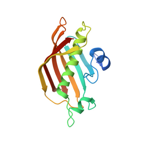
3AZB

Beta-Hydroxyacyl-Acyl Carrier Protein Dehydratase (FabZ) from Plasmodium falciparum in complex with NAS91-11


Experimental Data Snapshot

  • Method: X-RAY DIFFRACTION
  • Resolution: 2.60 Å
  • R-Value Free: 0.264 
  • R-Value Work: 0.226 
  • R-Value Observed: 0.228 

Starting Model: experimental
View more details

wwPDB Validation   3D Report Full Report


Ligand Structure Quality Assessment 


This is version 1.1 of the entry. See complete history


Literature

Structural basis for the functional and inhibitory mechanisms of beta-hydroxyacyl-acyl carrier protein dehydratase (FabZ) of Plasmodium falciparum

Maity, K.Venkata, B.S.Kapoor, N.Surolia, N.Surolia, A.Suguna, K.

(2011) J Struct Biol 176: 238-249

  • DOI: https://doi.org/10.1016/j.jsb.2011.07.018
  • Primary Citation of Related Structures:  
    3AZ8, 3AZ9, 3AZA, 3AZB

  • PubMed Abstract: 

    The β-hydroxyacyl-acyl carrier protein dehydratase of Plasmodium falciparum (PfFabZ) catalyzes the third and important reaction of the fatty acid elongation cycle. The crystal structure of PfFabZ is available in hexameric (active) and dimeric (inactive) forms. However, PfFabZ has not been crystallized with any bound inhibitors until now. We have designed a new condition to crystallize PfFabZ with its inhibitors bound in the active site, and determined the crystal structures of four of these complexes. This is the first report on any FabZ enzyme with active site inhibitors that interact directly with the catalytic residues. Inhibitor binding not only stabilized the substrate binding loop but also revealed that the substrate binding tunnel has an overall shape of "U". In the crystal structures, residue Phe169 located in the middle of the tunnel was found to be in two different conformations, open and closed. Thus, Phe169, merely by changing its side chain conformation, appears to be controlling the length of the tunnel to make it suitable for accommodating longer substrates. The volume of the substrate binding tunnel is determined by the sequence as well as by the conformation of the substrate binding loop region and varies between organisms for accommodating fatty acids of different chain lengths. This report on the crystal structures of the complexes of PfFabZ provides the structural basis of the inhibitory mechanism of the enzyme that could be used to improve the potency of inhibitors against an important component of fatty acid synthesis common to many infectious organisms.


  • Organizational Affiliation

    Molecular Biophysics Unit, Indian Institute of Science, Bangalore - 560 012, India.


Macromolecules
Find similar proteins by:  (by identity cutoff)  |  3D Structure
Entity ID: 1
MoleculeChains Sequence LengthOrganismDetailsImage
Beta-hydroxyacyl-ACP dehydratase
A, B, C, D, E
A, B, C, D, E, F, G, H, I, J, K, L, M, N, O, P, Q, R, S, T, U, V, W, X
154Plasmodium falciparumMutation(s): 0 
Gene Names: fabz
EC: 4.2.1 (PDB Primary Data), 4.2.1.59 (UniProt)
UniProt
Find proteins for Q965D7 (Plasmodium falciparum)
Explore Q965D7 
Go to UniProtKB:  Q965D7
Entity Groups  
Sequence Clusters30% Identity50% Identity70% Identity90% Identity95% Identity100% Identity
UniProt GroupQ965D7
Sequence Annotations
Expand
  • Reference Sequence
Small Molecules
Ligands 2 Unique
IDChains Name / Formula / InChI Key2D Diagram3D Interactions
KM1
Query on KM1

Download Ideal Coordinates CCD File 
CA [auth G],
EA [auth H],
GA [auth J],
KA [auth P],
Z [auth D]
5-chloro-8-[(3-chlorobenzyl)oxy]quinoline
C16 H11 Cl2 N O
BIHPUIBBWFPTCY-UHFFFAOYSA-N
GOL
Query on GOL

Download Ideal Coordinates CCD File 
AA [auth D]
BA [auth E]
DA [auth G]
FA [auth I]
HA [auth K]
AA [auth D],
BA [auth E],
DA [auth G],
FA [auth I],
HA [auth K],
IA [auth M],
JA [auth O],
LA [auth Q],
MA [auth S],
NA [auth V],
OA [auth X],
Y [auth A]
GLYCEROL
C3 H8 O3
PEDCQBHIVMGVHV-UHFFFAOYSA-N
Binding Affinity Annotations 
IDSourceBinding Affinity
KM1 PDBBind:  3AZB IC50: 1.20e+4 (nM) from 1 assay(s)
Experimental Data & Validation

Experimental Data

  • Method: X-RAY DIFFRACTION
  • Resolution: 2.60 Å
  • R-Value Free: 0.264 
  • R-Value Work: 0.226 
  • R-Value Observed: 0.228 
  • Space Group: I 41
Unit Cell:
Length ( Å )Angle ( ˚ )
a = 217.05α = 90
b = 217.05β = 90
c = 157.33γ = 90
Software Package:
Software NamePurpose
PHASERphasing
REFMACrefinement
MOSFLMdata reduction
SCALAdata scaling

Structure Validation

View Full Validation Report


Ligand Structure Quality Assessment 


Entry History 

Deposition Data

Revision History  (Full details and data files)

  • Version 1.0: 2012-02-29
    Type: Initial release
  • Version 1.1: 2023-11-01
    Changes: Data collection, Database references, Derived calculations, Refinement description