3AIA | pdb_00003aia

Crystal structure of DUF358 reveals a putative SPOUT-class methltransferase


Experimental Data Snapshot

  • Method: X-RAY DIFFRACTION
  • Resolution: 1.40 Å
  • R-Value Free: 
    0.218 (Depositor), 0.226 (DCC) 
  • R-Value Work: 
    0.188 (Depositor), 0.199 (DCC) 
  • R-Value Observed: 
    0.190 (Depositor) 

Starting Model: experimental
View more details

wwPDB Validation 3D Report Full Report

Validation slider image for 3AIA

Ligand Structure Quality Assessment 


This is version 1.2 of the entry. See complete history

Literature

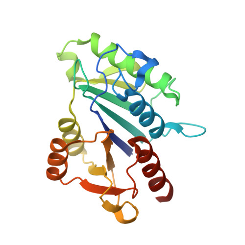
Crystal structure of Mj1640/DUF358 protein reveals a putative SPOUT-class RNA methyltransferase

Chen, H.Y.Yuan, Y.A.

(2010) J Mol Cell Biol 2: 366-374

  • DOI: https://doi.org/10.1093/jmcb/mjq034
  • Primary Citation Related Structures: 
    3AI9, 3AIA

  • PubMed Abstract: 

    The proteins in DUF358 family are all bacterial proteins, which are ∼200 amino acids long with unknown function. Bioinformatics analysis suggests that these proteins contain several conserved arginines and aspartates that might adopt SPOUT-class fold. Here we report crystal structure of Methanocaldococcus jannaschii DUF358/Mj1640 in complex with S-adenosyl-L-methionine (SAM) at 1.4 Å resolution. The structure reveals a single domain structure consisting of eight-stranded β-sheets sandwiched by six α-helices at both sides. Similar to other SPOUT-class members, Mj1640 contains a typical deep trefoil knot at its C-terminus to accommodate the SAM cofactor. However, Mj1640 has limited structural extension at its N-terminus, which is unique to this family member. Mj1640 forms a dimer, which is mediated by two parallel pairs of α-helices oriented almost perpendicular to each other. Although Mj1640 shares close structural similarity with Nep1, the significant differences in N-terminal extension domain and the overall surface charge distribution strongly suggest that Mj1640 might target a different RNA sequence. Detailed structural analysis of the SAM-binding pocket reveals that Asp157 or Glu183 from its own monomer or Ser43 from the associate monomer probably plays the catalytic role for RNA methylation.


  • Organizational Affiliation
    • Mechanobiology Institute, National University of Singapore, T-lab Building, 5A Engineering Drive 1, Singapore 117411, Singapore.

Macromolecule Content 

  • Total Structure Weight: 49.69 kDa 
  • Atom Count: 3,798 
  • Modeled Residue Count: 400 
  • Deposited Residue Count: 422 
  • Unique protein chains: 1

Macromolecules

Find similar proteins by:|  3D Structure
Entity ID: 1
MoleculeChains  Sequence LengthOrganismDetailsImage
UPF0217 protein MJ1640
A, B
211Methanocaldococcus jannaschiiMutation(s): 0 
Gene Names: MJ1640
EC: 2.1.1.257
UniProt
Find proteins for Q59034 (Methanocaldococcus jannaschii (strain ATCC 43067 / DSM 2661 / JAL-1 / JCM 10045 / NBRC 100440))
Explore Q59034 
Go to UniProtKB:  Q59034
Entity Groups
Sequence Clusters30% Identity50% Identity70% Identity90% Identity95% Identity100% Identity
UniProt GroupQ59034
Sequence Annotations
Expand
Reference Sequence

Experimental Data & Validation

Experimental Data

  • Method: X-RAY DIFFRACTION
  • Resolution: 1.40 Å
  • R-Value Free:  0.218 (Depositor), 0.226 (DCC) 
  • R-Value Work:  0.188 (Depositor), 0.199 (DCC) 
  • R-Value Observed: 0.190 (Depositor) 
Space Group: P 1 21 1
Unit Cell:
Length ( Å )Angle ( ˚ )
a = 42.862α = 90
b = 93.869β = 100.14
c = 53.837γ = 90
Software Package:
Software NamePurpose
HKL-2000data collection
MOLREPphasing
REFMACrefinement
HKL-2000data reduction
HKL-2000data scaling

Structure Validation

View Full Validation Report


Ligand Structure Quality Assessment 


Entry History 

Deposition Data

Revision History  (Full details and data files)

  • Version 1.0: 2011-03-30
    Type: Initial release
  • Version 1.1: 2011-07-13
    Changes: Version format compliance
  • Version 1.2: 2023-11-01
    Changes: Data collection, Database references, Derived calculations, Refinement description