31VJ | pdb_000031vj

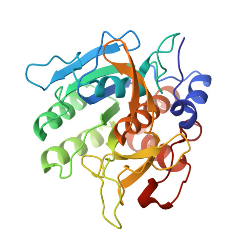
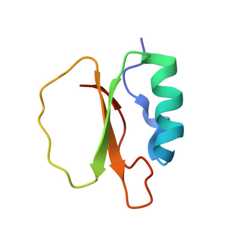
Alcalase/CI-2A(M59P) complex at 100K


Experimental Data Snapshot

  • Method: X-RAY DIFFRACTION
  • Resolution: 0.74 Å
  • R-Value Work: 
    0.095 (Depositor), 0.086 (DCC) 

Starting Model: experimental
View more details

wwPDB Validation 3D Report Full Report

Validation slider image for 31VJ

This is version 1.0 of the entry. See complete history. 

Literature

Catalytic hydrogen bonds in alcalase-CI2A complexes revealed by ultra-high-resolution crystallography.

Raczynska, J.E., Jedrzejczak, R., Dauter, M., Ostergaard, P.R., Wilson, K.S., Rypniewski, W.

(2026) J Struct Biol : 108377-108377

  • DOI: https://doi.org/10.1016/j.jsb.2026.108377
  • Primary Citation Related Structures: 
    31VH, 31VI, 31VJ

  • PubMed Abstract: 

    Serine proteases catalyze peptide-bond hydrolysis through a conserved catalytic triad and an extensive network of hydrogen bonds that facilitate proton transfer and stabilization of reaction intermediates. Despite decades of study, the nature of the hydrogen-bonding interactions within the catalytic machinery remains incompletely understood. Here, we report ultra-high-resolution crystal structures of alcalase, a subtilisin-family serine protease, in complex with native chymotrypsin inhibitor 2A (CI-2 A) and with a low-affinity M59P inhibitor variant. The cryogenic structures were determined at 0.74 Å resolution, while an additional 4 °C (277 K) structure of the native complex was determined at 1.05 Å resolution. The data allowed visualization and refinement of numerous hydrogen atoms within the active site and at the enzyme-inhibitor interface. Comparison of the 4 °C and cryogenic structures revealed a systematic shortening of hydrogen-bond donor-acceptor distances upon cooling, consistent with the global lattice contraction. A notable exception was the hydrogen bond between the catalytic residues Ser221 and His64, which remained unusually short at both temperatures. In the 4 °C structure, the corresponding hydrogen atom is associated with weak and diffuse electron density, suggesting increased proton mobility within this interaction. The structures further reveal temperature-dependent differences in interactions involving the scissile peptide bond of the inhibitor and the conserved backbone carbonyl oxygen of Ser125. These observations support a model in which Ser125 participates in a concerted hydrogen-bond network coupled to proton transfer within the catalytic triad. Together, the results provide atomic-level insights into hydrogen bonding, protonation equilibria, and catalytic mechanism in subtilisin-like serine proteases.


  • Organizational Affiliation: 
    • Institute of Bioorganic Chemistry, Polish Academy of Sciences, Noskowskiego 12/14, 61-704 Poznan, Poland.

Macromolecule Content 

  • Total Structure Weight: 34.95 kDa 
  • Atom Count: 3,074 
  • Modeled Residue Count: 338 
  • Deposited Residue Count: 338 
  • Unique protein chains: 2

Macromolecules

Find similar proteins by:|  3D Structure
Entity ID: 1
MoleculeChains  Sequence LengthOrganismDetailsImage
KerA274Bacillus licheniformisMutation(s): 0 
EC: 3.4
UniProt
Find proteins for Q9FDF2 (Bacillus licheniformis)
Explore Q9FDF2 
Go to UniProtKB:  Q9FDF2
Entity Groups
Sequence Clusters30% Identity50% Identity70% Identity90% Identity95% Identity100% Identity
UniProt GroupQ9FDF2
Sequence Annotations
Expand
Reference Sequence
Find similar proteins by:|  3D Structure
Entity ID: 2
MoleculeChains  Sequence LengthOrganismDetailsImage
Subtilisin-chymotrypsin inhibitor-2A64Hordeum vulgareMutation(s): 0 
UniProt
Find proteins for P01053 (Hordeum vulgare)
Explore P01053 
Go to UniProtKB:  P01053
Entity Groups
Sequence Clusters30% Identity50% Identity70% Identity90% Identity95% Identity100% Identity
UniProt GroupP01053
Sequence Annotations
Expand
Reference Sequence

Small Molecules

Ligands 4 Unique
IDChains Name / Formula / InChI Key2D Diagram3D Interactions
GOL

Query on GOL


Download:Ideal Coordinates CCD File
I [auth A],
J [auth A]
GLYCEROL
C3 H8 O3
PEDCQBHIVMGVHV-UHFFFAOYSA-N
CA

Query on CA


Download:Ideal Coordinates CCD File
C [auth A]CALCIUM ION
Ca
BHPQYMZQTOCNFJ-UHFFFAOYSA-N
CL

Query on CL


Download:Ideal Coordinates CCD File
G [auth A],
H [auth A]
CHLORIDE ION
Cl
VEXZGXHMUGYJMC-UHFFFAOYSA-M
NA

Query on NA


Download:Ideal Coordinates CCD File
D [auth A],
E [auth A],
F [auth A]
SODIUM ION
Na
FKNQFGJONOIPTF-UHFFFAOYSA-N

Experimental Data & Validation

Experimental Data

  • Method: X-RAY DIFFRACTION
  • Resolution: 0.74 Å
  • R-Value Work:  0.095 (Depositor), 0.086 (DCC) 
Space Group: P 1
Unit Cell:
Length ( Å )Angle ( ˚ )
a = 37.78α = 103.7
b = 44.52β = 101.3
c = 46.18γ = 103.2
Software Package:
Software NamePurpose
SHELXrefinement
SCALEPACKdata scaling
DENZOdata reduction
AMoREphasing
PDB_EXTRACTdata extraction

Structure Validation

View Full Validation Report


Entry History 

& Funding Information

Deposition Data


Funding OrganizationLocationGrant Number
Not funded--

Revision History  (Full details and data files)

  • Version 1.0: 2026-10-07
    Type: Initial release