


Crystal structure of uncharacterized protein Tfu_2867 (YP_290923.1) from Thermobifida fusca YX at 1.95 A resolution
Joint Center for Structural Genomics (JCSG)To be published.
Experimental Data Snapshot
Entity ID: 1 | |||||
|---|---|---|---|---|---|
| Molecule | Chains | Sequence Length | Organism | Details | Image |
| Uncharacterized protein Tfu_2867 | 274 | Thermobifida fusca YX | Mutation(s): 1 Gene Names: YP_290923.1, Tfu_2867 | 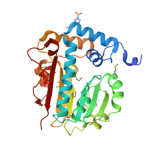 | |
UniProt | |||||
Entity Groups | |||||
| Sequence Clusters | 30% Identity50% Identity70% Identity90% Identity95% Identity100% Identity | ||||
| UniProt Group | Q47KX2 | ||||
Sequence AnnotationsExpand | |||||
Reference Sequence | |||||
| Ligands 3 Unique | |||||
|---|---|---|---|---|---|
| ID | Chains | Name / Formula / InChI Key | 2D Diagram | 3D Interactions | |
| SAM Download:Ideal Coordinates CCD File | F [auth A], O [auth B] | S-ADENOSYLMETHIONINE C15 H22 N6 O5 S MEFKEPWMEQBLKI-FCKMPRQPSA-N |  | ||
| MPD Download:Ideal Coordinates CCD File | G [auth A] H [auth A] I [auth A] P [auth B] Q [auth B] | (4S)-2-METHYL-2,4-PENTANEDIOL C6 H14 O2 SVTBMSDMJJWYQN-YFKPBYRVSA-N |  | ||
| SO4 Download:Ideal Coordinates CCD File | C [auth A] D [auth A] E [auth A] J [auth B] K [auth B] | SULFATE ION O4 S QAOWNCQODCNURD-UHFFFAOYSA-L |  | ||
| Modified Residues 2 Unique | |||||
|---|---|---|---|---|---|
| ID | Chains | Type | Formula | 2D Diagram | Parent |
| MSE Query on MSE | A, B | L-PEPTIDE LINKING | C5 H11 N O2 Se |  | MET |
| NEP Query on NEP | A, B | L-PEPTIDE LINKING | C6 H10 N3 O5 P |  | HIS |
| Length ( Å ) | Angle ( ˚ ) |
|---|---|
| a = 102.627 | α = 90 |
| b = 76.024 | β = 90 |
| c = 81.315 | γ = 90 |
| Software Name | Purpose |
|---|---|
| REFMAC | refinement |
| PHENIX | refinement |
| SHELX | phasing |
| MolProbity | model building |
| SCALA | data scaling |
| PDB_EXTRACT | data extraction |
| MAR345 | data collection |
| MOSFLM | data reduction |
| SHELXD | phasing |
| autoSHARP | phasing |


