2FVY | pdb_00002fvy

High Resolution Glucose Bound Crystal Structure of GGBP


Experimental Data Snapshot

  • Method: X-RAY DIFFRACTION
  • Resolution: 0.92 Å
  • R-Value Free: 
    0.130 (Depositor), 0.105 (DCC) 
  • R-Value Work: 
    0.110 (Depositor) 
  • R-Value Observed: 
    0.118 (Depositor) 

Starting Model: experimental
View more details

wwPDB Validation 3D Report Full Report

Validation slider image for 2FVY

Ligand Structure Quality Assessment 


This is version 2.4 of the entry. See complete history

Literature

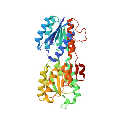
Conformational changes of glucose/galactose-binding protein illuminated by open, unliganded, and ultra-high-resolution ligand-bound structures.

Borrok, M.J.Kiessling, L.L.Forest, K.T.

(2007) Protein Sci 16: 1032-1041

  • DOI: https://doi.org/10.1110/ps.062707807
  • Primary Citation Related Structures: 
    2FVY, 2FW0

  • PubMed Abstract: 

    D-Glucose/D-Galactose-binding protein (GGBP) mediates chemotaxis toward and active transport of glucose and galactose in a number of bacterial species. GGBP, like other periplasmic binding proteins, can exist in open (ligand-free) and closed (ligand-bound) states. We report a 0.92 angstroms resolution structure of GGBP from Escherichia coli in the glucose-bound state and the first structure of an open, unbound form of GGBP (at 1.55 angstroms resolution). These structures vary in the angle between the two structural domains; the observed difference of 31 degrees arises from torsion angle changes in a three-segment hinge. A comparison with the closely related periplasmic receptors, ribose- and allose-binding proteins, shows that the GGBP hinge residue positions that undergo the largest conformational changes are different. Furthermore, the high-quality data collected for the atomic resolution glucose-bound structure allow for the refinement of specific hydrogen atom positions, the assignment of alternate side chain conformations, the first description of CO(2) trapped after radiation-induced decarboxylation, and insight into the role of the exo-anomeric effect in sugar binding. Together, these structures provide insight into how the hinge-bending movement of GGBP facilitates ligand binding, transport, and signaling.


  • Organizational Affiliation
    • Department of Biochemistry, University of Wisconsin, Madison, Wisconsin 53706, USA.

Macromolecule Content 

  • Total Structure Weight: 33.82 kDa 
  • Atom Count: 2,837 
  • Modeled Residue Count: 305 
  • Deposited Residue Count: 309 
  • Unique protein chains: 1

Macromolecules

Find similar proteins by:|  3D Structure
Entity ID: 1
MoleculeChains  Sequence LengthOrganismDetailsImage
D-galactose-binding periplasmic protein309Escherichia coliMutation(s): 0 
Gene Names: mglB
UniProt
Find proteins for P0AEE5 (Escherichia coli (strain K12))
Explore P0AEE5 
Go to UniProtKB:  P0AEE5
Entity Groups
Sequence Clusters30% Identity50% Identity70% Identity90% Identity95% Identity100% Identity
UniProt GroupP0AEE5
Sequence Annotations
Expand
Reference Sequence

Small Molecules

Ligands 5 Unique
IDChains Name / Formula / InChI Key2D Diagram3D Interactions
BGC

Query on BGC


Download:Ideal Coordinates CCD File
B [auth A]beta-D-glucopyranose
C6 H12 O6
WQZGKKKJIJFFOK-VFUOTHLCSA-N
GOL

Query on GOL


Download:Ideal Coordinates CCD File
F [auth A]GLYCEROL
C3 H8 O3
PEDCQBHIVMGVHV-UHFFFAOYSA-N
ACT

Query on ACT


Download:Ideal Coordinates CCD File
D [auth A]ACETATE ION
C2 H3 O2
QTBSBXVTEAMEQO-UHFFFAOYSA-M
CO2

Query on CO2


Download:Ideal Coordinates CCD File
E [auth A]CARBON DIOXIDE
C O2
CURLTUGMZLYLDI-UHFFFAOYSA-N
CA

Query on CA


Download:Ideal Coordinates CCD File
C [auth A]CALCIUM ION
Ca
BHPQYMZQTOCNFJ-UHFFFAOYSA-N

Experimental Data & Validation

Experimental Data

  • Method: X-RAY DIFFRACTION
  • Resolution: 0.92 Å
  • R-Value Free:  0.130 (Depositor), 0.105 (DCC) 
  • R-Value Work:  0.110 (Depositor) 
  • R-Value Observed: 0.118 (Depositor) 
Space Group: C 1 2 1
Unit Cell:
Length ( Å )Angle ( ˚ )
a = 119.989α = 90
b = 36.238β = 124.5
c = 80.101γ = 90
Software Package:
Software NamePurpose
SHELXL-97refinement
SCALEPACKdata scaling
AMoREphasing
CNSrefinement
HKL-2000data collection
HKL-2000data reduction

Structure Validation

View Full Validation Report


Ligand Structure Quality Assessment 


Entry History 

Deposition Data

Revision History  (Full details and data files)

  • Version 1.0: 2007-02-06
    Type: Initial release
  • Version 1.1: 2008-05-01
    Changes: Version format compliance
  • Version 1.2: 2011-07-13
    Changes: Atomic model, Version format compliance
  • Version 1.3: 2019-07-24
    Changes: Data collection, Derived calculations, Refinement description
  • Version 2.0: 2020-07-29
    Type: Remediation
    Reason: Carbohydrate remediation
    Changes: Atomic model, Data collection, Derived calculations, Structure summary
  • Version 2.1: 2020-09-02
    Changes: Structure summary
  • Version 2.2: 2020-10-21
    Changes: Derived calculations
  • Version 2.3: 2024-02-14
    Changes: Data collection, Database references
  • Version 2.4: 2024-04-03
    Changes: Refinement description