2ZOI | pdb_00002zoi

Neutron Crystal Structure of Photoactive Yellow Protein, Wild type, at 295K


Experimental Data Snapshot

  • Method: NEUTRON DIFFRACTION
  • Resolution: 1.50 Å
  • R-Value Free: 
    0.219 (Depositor) 
  • R-Value Work: 
    0.192 (Depositor) 

Starting Model: experimental
View more details

wwPDB Validation 3D Report Full Report

Validation slider image for 2ZOI

This is version 1.3 of the entry. See complete history

Literature

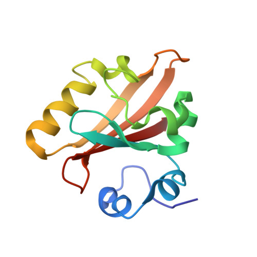
Low-barrier hydrogen bond in photoactive yellow protein

Yamaguchi, S.Kamikubo, H.Kurihara, K.Kuroki, R.Niimura, N.Shimizu, N.Yamazaki, Y.Kataoka, M.

(2009) Proc Natl Acad Sci U S A 106: 440-444

  • DOI: https://doi.org/10.1073/pnas.0811882106
  • Primary Citation Related Structures: 
    2ZOH, 2ZOI

  • PubMed Abstract: 

    Low-barrier hydrogen bonds (LBHBs) have been proposed to play roles in protein functions, including enzymatic catalysis and proton transfer. Transient formation of LBHBs is expected to stabilize specific reaction intermediates. However, based on experimental results and theoretical considerations, arguments against the importance of LBHB in proteins have been raised. The discrepancy is caused by the absence of direct identification of the hydrogen atom position. Here, we show by high-resolution neutron crystallography of photoactive yellow protein (PYP) that a LBHB exists in a protein, even in the ground state. We identified approximately 87% (819/942) of the hydrogen positions in PYP and demonstrated that the hydrogen bond between the chromophore and E46 is a LBHB. This LBHB stabilizes an isolated electric charge buried in the hydrophobic environment of the protein interior. We propose that in the excited state the fast relaxation of the LBHB into a normal hydrogen bond is the trigger for photo-signal propagation to the protein moiety. These results give insights into the novel roles of LBHBs and the mechanism of the formation of LBHBs.


  • Organizational Affiliation
    • Graduate School of Materials Science, Nara Institute of Science and Technology, 8916-5 Takayama, Ikoma, Nara 630-0192, Japan.

Macromolecule Content 

  • Total Structure Weight: 14.05 kDa 
  • Atom Count: 1,094 
  • Modeled Residue Count: 125 
  • Deposited Residue Count: 125 
  • Unique protein chains: 1

Macromolecules

Find similar proteins by:|  3D Structure
Entity ID: 1
MoleculeChains  Sequence LengthOrganismDetailsImage
Photoactive yellow protein125Halorhodospira halophilaMutation(s): 0 
Gene Names: PYP
UniProt
Find proteins for P16113 (Halorhodospira halophila)
Explore P16113 
Go to UniProtKB:  P16113
Entity Groups
Sequence Clusters30% Identity50% Identity70% Identity90% Identity95% Identity100% Identity
UniProt GroupP16113
Sequence Annotations
Expand
Reference Sequence

Small Molecules

Ligands 1 Unique
IDChains Name / Formula / InChI Key2D Diagram3D Interactions
HC4

Query on HC4


Download:Ideal Coordinates CCD File
B [auth A]4'-HYDROXYCINNAMIC ACID
C9 H8 O3
NGSWKAQJJWESNS-ZZXKWVIFSA-N

Experimental Data & Validation

Experimental Data

  • Method: NEUTRON DIFFRACTION
  • Resolution: 1.50 Å
  • R-Value Free:  0.219 (Depositor) 
  • R-Value Work:  0.192 (Depositor) 
Space Group: P 63
Unit Cell:
Length ( Å )Angle ( ˚ )
a = 67.145α = 90
b = 67.145β = 90
c = 41.119γ = 120
Software Package:
Software NamePurpose
AMoREphasing
CNSrefinement
MACSCIENCEdata collection
DENZOdata reduction
SCALEPACKdata scaling

Structure Validation

View Full Validation Report


Entry History 

Deposition Data

Revision History  (Full details and data files)

  • Version 1.0: 2009-03-24
    Type: Initial release
  • Version 1.1: 2011-07-13
    Changes: Non-polymer description, Version format compliance
  • Version 1.2: 2018-03-07
    Changes: Data collection
  • Version 1.3: 2023-11-01
    Changes: Data collection, Database references, Derived calculations, Refinement description