2YKO | pdb_00002yko

Structure of the human LINE-1 ORF1p trimer


Experimental Data Snapshot

  • Method: X-RAY DIFFRACTION
  • Resolution: 2.10 Å
  • R-Value Free: 
    0.264 (Depositor), 0.263 (DCC) 
  • R-Value Work: 
    0.208 (Depositor), 0.207 (DCC) 
  • R-Value Observed: 
    0.211 (Depositor) 

Starting Models: experimental
View more details

wwPDB Validation 3D Report Full Report

Validation slider image for 2YKO

This is version 1.5 of the entry. See complete history

Literature

Trimeric Structure and Flexibility of the L1Orf1P Protein in Human L1 Retrotransposition

Khazina, E.Truffault, V.Buettner, R.Schmidt, S.Coles, M.Weichenrieder, O.

(2011) Nat Struct Mol Biol 18: 1006

  • DOI: https://doi.org/10.1038/nsmb.2097
  • Primary Citation Related Structures: 
    2LDY, 2YKO, 2YKP, 2YKQ

  • PubMed Abstract: 

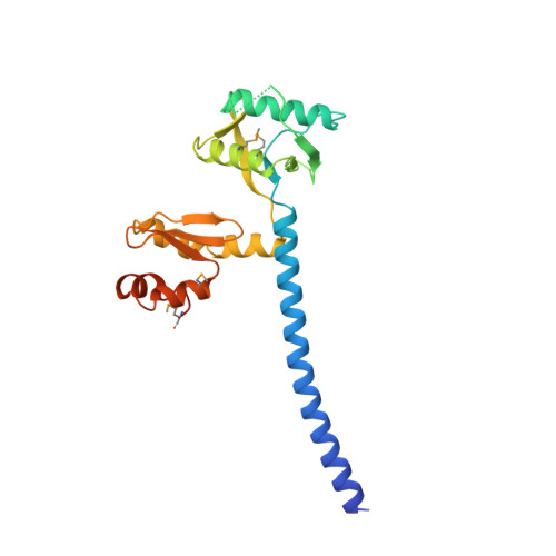
    The LINE-1 (L1) retrotransposon emerges as a major source of human interindividual genetic variation, with important implications for evolution and disease. L1 retrotransposition is poorly understood at the molecular level, and the mechanistic details and evolutionary origin of the L1-encoded L1ORF1 protein (L1ORF1p) are particularly obscure. Here three crystal structures of trimeric L1ORF1p and NMR solution structures of individual domains reveal a sophisticated and highly structured, yet remarkably flexible, RNA-packaging protein. It trimerizes via an N-terminal, ion-containing coiled coil that serves as scaffold for the flexible attachment of the central RRM and the C-terminal CTD domains. The structures explain the specificity for single-stranded RNA substrates, and a mutational analysis indicates that the precise control of domain flexibility is critical for retrotransposition. Although the evolutionary origin of L1ORF1p remains unclear, our data reveal previously undetected structural and functional parallels to viral proteins.


  • Organizational Affiliation
    • Department of Biochemistry, Max Planck Institute for Developmental Biology, Tübingen, Germany.

Macromolecule Content 

  • Total Structure Weight: 84.14 kDa 
  • Atom Count: 5,206 
  • Modeled Residue Count: 598 
  • Deposited Residue Count: 699 
  • Unique protein chains: 1

Macromolecules

Find similar proteins by:|  3D Structure
Entity ID: 1
MoleculeChains  Sequence LengthOrganismDetailsImage
LINE-1 ORF1P
A, B, C
233Homo sapiensMutation(s): 5 
UniProt
Find proteins for Q9UN81 (Homo sapiens)
Explore Q9UN81 
Go to UniProtKB:  Q9UN81
Entity Groups
Sequence Clusters30% Identity50% Identity70% Identity90% Identity95% Identity100% Identity
UniProt GroupQ9UN81
Sequence Annotations
Expand
Reference Sequence

Small Molecules

Modified Residues  1 Unique
IDChains TypeFormula2D DiagramParent
MSE
Query on MSE
A, B, C
L-PEPTIDE LINKINGC5 H11 N O2 SeMET

Experimental Data & Validation

Experimental Data

  • Method: X-RAY DIFFRACTION
  • Resolution: 2.10 Å
  • R-Value Free:  0.264 (Depositor), 0.263 (DCC) 
  • R-Value Work:  0.208 (Depositor), 0.207 (DCC) 
  • R-Value Observed: 0.211 (Depositor) 
Space Group: P 21 21 21
Unit Cell:
Length ( Å )Angle ( ˚ )
a = 72.69α = 90
b = 85.42β = 90
c = 111.45γ = 90
Software Package:
Software NamePurpose
XDSdata reduction
XSCALEdata scaling
SHELXphasing
PHASERphasing
PHENIXrefinement

Structure Validation

View Full Validation Report


Entry History 

Deposition Data

Revision History  (Full details and data files)

  • Version 1.0: 2011-08-10
    Type: Initial release
  • Version 1.1: 2011-08-24
    Changes: Atomic model, Database references, Derived calculations, Other
  • Version 1.2: 2011-09-14
    Changes: Database references
  • Version 1.3: 2012-10-10
    Changes: Database references
  • Version 1.4: 2023-12-20
    Changes: Data collection, Database references, Derived calculations, Other, Refinement description
  • Version 1.5: 2024-11-20
    Changes: Structure summary