2YGQ | pdb_00002ygq

WIF domain-epidermal growth factor (EGF)-like domains 1-3 of human Wnt inhibitory factor 1 in complex with 1,2- dipalmitoylphosphatidylcholine


Experimental Data Snapshot

  • Method: X-RAY DIFFRACTION
  • Resolution: 3.95 Å
  • R-Value Free: 
    0.331 (Depositor), 0.340 (DCC) 
  • R-Value Work: 
    0.297 (Depositor), 0.299 (DCC) 
  • R-Value Observed: 
    0.298 (Depositor) 

wwPDB Validation 3D Report Full Report

Validation slider image for 2YGQ

Ligand Structure Quality Assessment 


This is version 2.1 of the entry. See complete history

Literature

Modular Mechanism of Wnt Signaling Inhibition by Wnt Inhibitory Factor 1

Malinauskas, T.Aricescu, A.R.Lu, W.Siebold, C.Jones, E.Y.

(2011) Nat Struct Mol Biol 18: 886

  • DOI: https://doi.org/10.1038/nsmb.2081
  • Primary Citation Related Structures: 
    2YGN, 2YGO, 2YGP, 2YGQ

  • PubMed Abstract: 

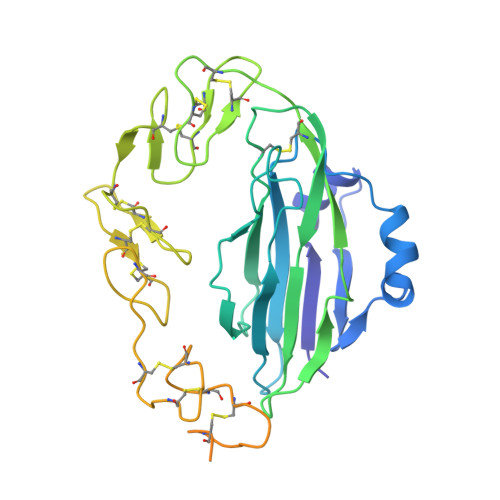
    Wnt morphogens control embryonic development and homeostasis in adult tissues. In vertebrates the N-terminal WIF domain (WIF-1(WD)) of Wnt inhibitory factor 1 (WIF-1) binds Wnt ligands. Our crystal structure of WIF-1(WD) reveals a previously unidentified binding site for phospholipid; two acyl chains extend deep into the domain, and the head group is exposed to the surface. Biophysical and cellular assays indicate that there is a WIF-1(WD) Wnt-binding surface proximal to the lipid head group but also implicate the five epidermal growth factor (EGF)-like domains (EGFs I-V) in Wnt binding. The six-domain WIF-1 crystal structure shows that EGFs I-V are wrapped back, interfacing with WIF-1(WD) at EGF III. EGFs II-V contain a heparan sulfate proteoglycan (HSPG)-binding site, consistent with conserved positively charged residues on EGF IV. This combination of HSPG- and Wnt-binding properties suggests a modular model for the localization of WIF-1 and for signal inhibition within morphogen gradients.


  • Organizational Affiliation
    • Division of Structural Biology, Wellcome Trust Centre for Human Genetics, University of Oxford, Oxford, UK.

Macromolecule Content 

  • Total Structure Weight: 37.73 kDa 
  • Atom Count: 1,969 
  • Modeled Residue Count: 241 
  • Deposited Residue Count: 324 
  • Unique protein chains: 1

Macromolecules

Find similar proteins by:|  3D Structure
Entity ID: 1
MoleculeChains  Sequence LengthOrganismDetailsImage
WNT INHIBITORY FACTOR 1324Homo sapiensMutation(s): 1 
UniProt & NIH Common Fund Data Resources
Find proteins for Q9Y5W5 (Homo sapiens)
Explore Q9Y5W5 
Go to UniProtKB:  Q9Y5W5
PHAROS:  Q9Y5W5
GTEx:  ENSG00000156076 
Entity Groups
Sequence Clusters30% Identity50% Identity70% Identity90% Identity95% Identity100% Identity
UniProt GroupQ9Y5W5
Glycosylation
Glycosylation Sites: 3Go to GlyGen: Q9Y5W5-1
Sequence Annotations
Expand
Reference Sequence

Oligosaccharides

Help  
Entity ID: 2
MoleculeChains Length2D Diagram GlycosylationD Interactions
1,3,4,6-tetra-O-sulfo-beta-D-fructofuranose-(2-1)-2,3,4,6-tetra-O-sulfonato-alpha-D-glucopyranose
B
2N/A
Glycosylation Resources
GlyTouCan: G97555GV
GlyCosmos: G97555GV

Biologically Interesting Molecules (External Reference) 

1 Unique

Experimental Data & Validation

Experimental Data

  • Method: X-RAY DIFFRACTION
  • Resolution: 3.95 Å
  • R-Value Free:  0.331 (Depositor), 0.340 (DCC) 
  • R-Value Work:  0.297 (Depositor), 0.299 (DCC) 
  • R-Value Observed: 0.298 (Depositor) 
Space Group: I 21 3
Unit Cell:
Length ( Å )Angle ( ˚ )
a = 178.007α = 90
b = 178.007β = 90
c = 178.007γ = 90
Software Package:
Software NamePurpose
PHENIXrefinement
DENZOdata reduction
SCALEPACKdata scaling
PHASERphasing

Structure Validation

View Full Validation Report


Ligand Structure Quality Assessment 


Entry History 

Deposition Data

Revision History  (Full details and data files)

  • Version 1.0: 2011-07-13
    Type: Initial release
  • Version 1.1: 2011-08-17
    Changes: Database references
  • Version 2.0: 2020-07-29
    Type: Remediation
    Reason: Carbohydrate remediation
    Changes: Atomic model, Data collection, Derived calculations, Non-polymer description, Other, Structure summary
  • Version 2.1: 2024-11-06
    Changes: Data collection, Database references, Structure summary