2XSM | pdb_00002xsm

Crystal structure of the mammalian cytosolic chaperonin CCT in complex with tubulin


Experimental Data Snapshot

  • Method: X-RAY DIFFRACTION
  • Resolution: 5.50 Å
  • R-Value Work: 
    0.416 (DCC) 

wwPDB Validation 3D Report Full Report

Validation slider image for 2XSM

This is version 1.1 of the entry. See complete history

Re-refinement Note

A newer entry is available that reflects an alternative modeling of the original data: 4B2T

Literature

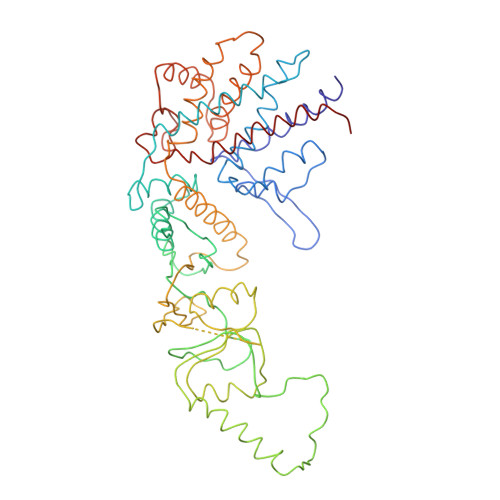
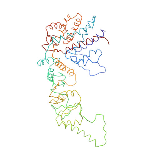
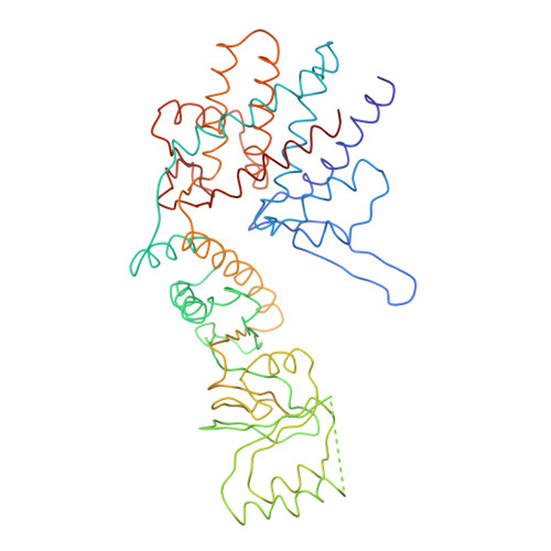
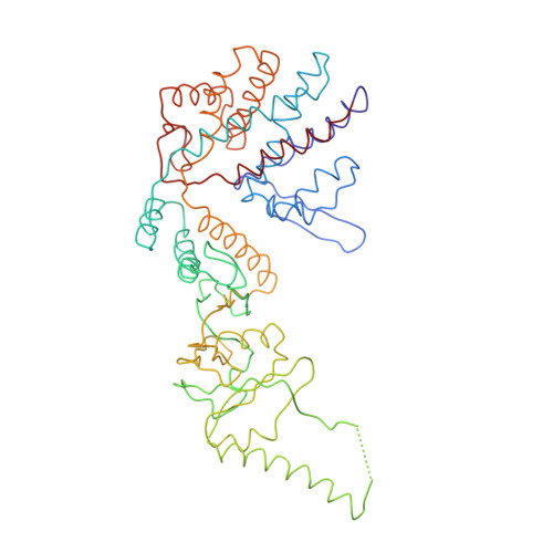
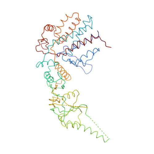
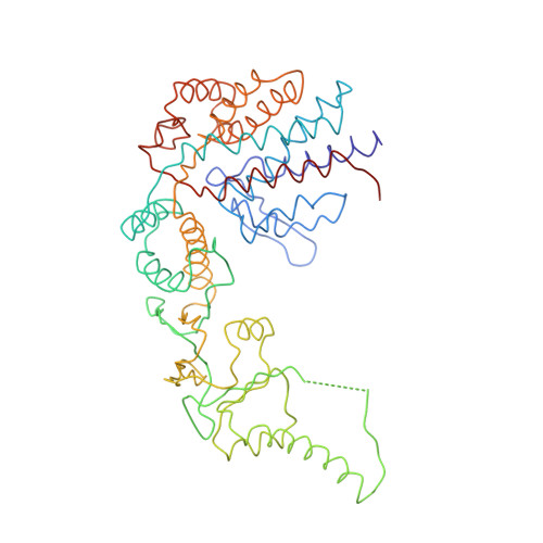
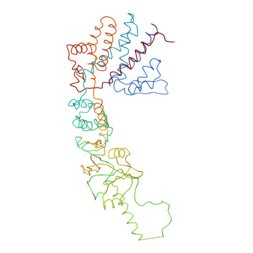
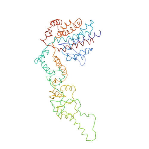
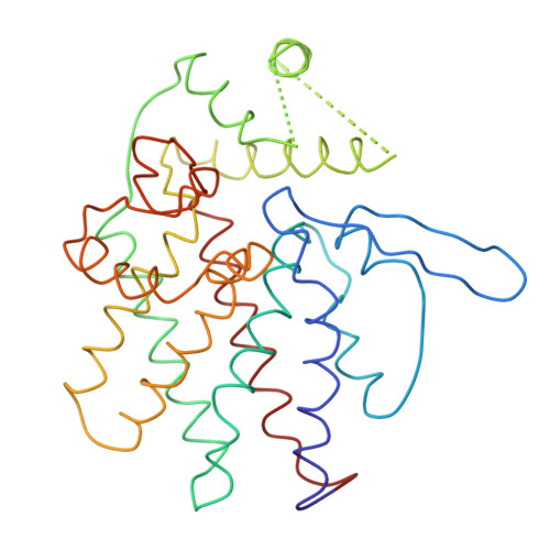
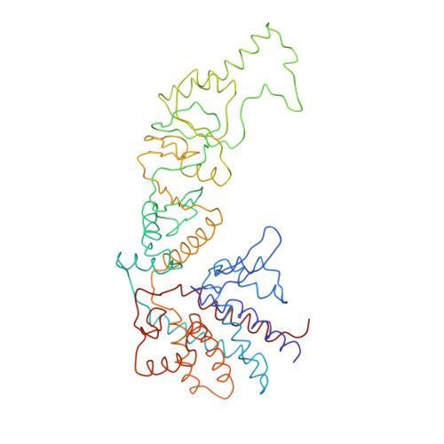
Crystal Structure of the Open Conformation of the Mammalian Chaperonin Cct in Complex with Tubulin.

Munoz, I.G.Yebenes, H.Zhou, M.Mesa, P.Serna, M.Park, A.Y.Bragado-Nilsson, E.Beloso, A.De Carcer, G.Malumbres, M.Robinson, C.V.Valpuesta, J.M.Montoya, G.

(2011) Nat Struct Mol Biol 18: 14

  • DOI: https://doi.org/10.1038/nsmb.1971
  • Primary Citation Related Structures: 
    2XSM

  • PubMed Abstract: 

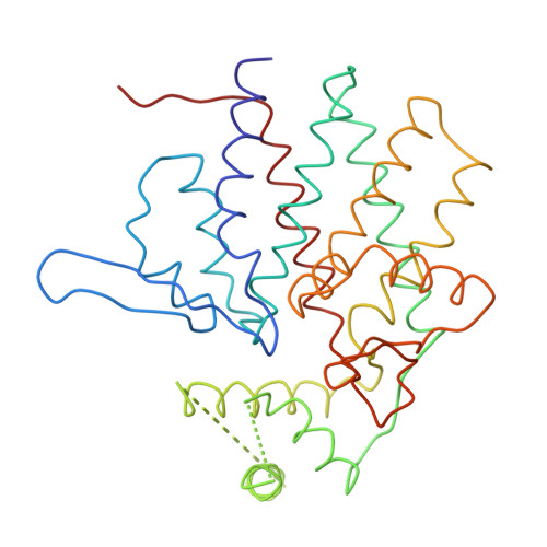
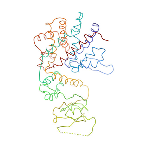
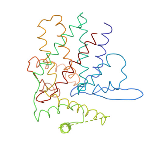
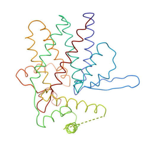
    Protein folding is assisted by molecular chaperones. CCT (chaperonin containing TCP-1, or TRiC) is a 1-MDa oligomer that is built by two rings comprising eight different 60-kDa subunits. This chaperonin regulates the folding of important proteins including actin, α-tubulin and β-tubulin. We used an electron density map at 5.5 Å resolution to reconstruct CCT, which showed a substrate in the inner cavities of both rings. Here we present the crystal structure of the open conformation of this nanomachine in complex with tubulin, providing information about the mechanism by which it aids tubulin folding. The structure showed that the substrate interacts with loops in the apical and equatorial domains of CCT. The organization of the ATP-binding pockets suggests that the substrate is stretched inside the cavity. Our data provide the basis for understanding the function of this chaperonin.


  • Organizational Affiliation
    • Macromolecular Crystallography Group, Structural Biology and Biocomputing Programme, Spanish National Cancer Research Centre (CNIO), Madrid, Spain.

Macromolecule Content 

  • Total Structure Weight: 548.19 kDa 
  • Atom Count: 6,438 
  • Modeled Residue Count: 6,438 
  • Deposited Residue Count: 6,438 
  • Unique protein chains: 15

Macromolecules

Find similar proteins by:|  3D Structure
Entity ID: 1
MoleculeChains  Sequence LengthOrganismDetailsImage
CCT489Bos taurusMutation(s): 0 
Find similar proteins by:|  3D Structure
Entity ID: 2
MoleculeChains  Sequence LengthOrganismDetailsImage
CCT478Bos taurusMutation(s): 0 
Find similar proteins by:|  3D Structure
Entity ID: 3
MoleculeChains  Sequence LengthOrganismDetailsImage
CCT455Bos taurusMutation(s): 0 
Find similar proteins by:|  3D Structure
Entity ID: 4
MoleculeChains  Sequence LengthOrganismDetailsImage
CCT471Bos taurusMutation(s): 0 
Find similar proteins by:|  3D Structure
Entity ID: 5
MoleculeChains  Sequence LengthOrganismDetailsImage
CCT472Bos taurusMutation(s): 0 
Find similar proteins by:|  3D Structure
Entity ID: 6
MoleculeChains  Sequence LengthOrganismDetailsImage
CCT466Bos taurusMutation(s): 0 
Find similar proteins by:|  3D Structure
Entity ID: 7
MoleculeChains  Sequence LengthOrganismDetailsImage
CCT485Bos taurusMutation(s): 0 
Find similar proteins by:|  3D Structure
Entity ID: 8
MoleculeChains  Sequence LengthOrganismDetailsImage
CCT474Bos taurusMutation(s): 0 
Find similar proteins by:|  3D Structure
Entity ID: 9
MoleculeChains  Sequence LengthOrganismDetailsImage
CCT293Bos taurusMutation(s): 0 
Find similar proteins by:|  3D Structure
Entity ID: 10
MoleculeChains  Sequence LengthOrganismDetailsImage
CCT299Bos taurusMutation(s): 0 
Find similar proteins by:|  3D Structure
Entity ID: 11
MoleculeChains  Sequence LengthOrganismDetailsImage
CCT394Bos taurusMutation(s): 0 
Find similar proteins by:|  3D Structure
Entity ID: 12
MoleculeChains  Sequence LengthOrganismDetailsImage
CCT
L, O
297Bos taurusMutation(s): 0 
Find similar proteins by:|  3D Structure
Entity ID: 13
MoleculeChains  Sequence LengthOrganismDetailsImage
CCT298Bos taurusMutation(s): 0 
Find similar proteins by:|  3D Structure
Entity ID: 14
MoleculeChains  Sequence LengthOrganismDetailsImage
CCT289Bos taurusMutation(s): 0 
Find similar proteins by:|  3D Structure
Entity ID: 15
MoleculeChains  Sequence LengthOrganismDetailsImage
CCT481Bos taurusMutation(s): 0 

Experimental Data & Validation

Experimental Data

  • Method: X-RAY DIFFRACTION
  • Resolution: 5.50 Å
  • R-Value Work:  0.416 (DCC) 
Space Group: P 21 21 2
Unit Cell:
Length ( Å )Angle ( ˚ )
a = 272.7α = 90
b = 313.5β = 90
c = 158.3γ = 90
Software Package:
Software NamePurpose
REFMACrefinement
XDSdata reduction
XSCALEdata scaling

Structure Validation

View Full Validation Report


Entry History 

Deposition Data

Revision History  (Full details and data files)

  • Version 1.0: 2010-12-15
    Type: Initial release
  • Version 1.1: 2024-05-08
    Changes: Database references, Other