2XRB | pdb_00002xrb

Structure of the N-terminal four domains of the complement regulator Rat Crry


Experimental Data Snapshot

  • Method: X-RAY DIFFRACTION
  • Resolution: 2.50 Å
  • R-Value Free: 
    0.237 (Depositor), 0.254 (DCC) 
  • R-Value Work: 
    0.185 (Depositor), 0.189 (DCC) 
  • R-Value Observed: 
    0.187 (Depositor) 

Starting Models: experimental
View more details

wwPDB Validation 3D Report Full Report

Validation slider image for 2XRB

This is version 1.3 of the entry. See complete history

Literature

Structures of the Rat Complement Regulator Crry.

Roversi, P.Johnson, S.Caesar, J.J.E.Mclean, F.Leath, K.J.Tsiftsoglou, S.A.Morgan, B.P.Harris, C.L.Sim, R.B.Lea, S.M.

(2011) Acta Crystallogr Sect F Struct Biol Cryst Commun 67: 739

  • DOI: https://doi.org/10.1107/S1744309111016551
  • Primary Citation Related Structures: 
    2XRB, 2XRD

  • PubMed Abstract: 

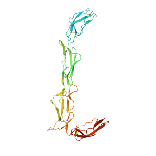
    Complement receptor 1-related protein Y (CrrY) is an important cell-surface regulator of complement that is unique to rodent species. The structure of rat CrrY domains 1-4 has been determined in two distinct crystal forms and reveals a 70° bend between domains 3 and 4. Comparisons of this structure with those of other complement regulators suggests that rearrangement of this interface may occur on forming the regulatory complex with C3b.


  • Organizational Affiliation
    • Sir William Dunn School of Pathology, Oxford University, South Parks Road, Oxford OX4 3RE, England.

Macromolecule Content 

  • Total Structure Weight: 33.02 kDa 
  • Atom Count: 2,158 
  • Modeled Residue Count: 254 
  • Deposited Residue Count: 290 
  • Unique protein chains: 1

Macromolecules

Find similar proteins by:|  3D Structure
Entity ID: 1
MoleculeChains  Sequence LengthOrganismDetailsImage
COMPLEMENT REGULATORY PROTEIN CRRY290Rattus norvegicusMutation(s): 0 
UniProt
Find proteins for Q63135 (Rattus norvegicus)
Explore Q63135 
Go to UniProtKB:  Q63135
Entity Groups
Sequence Clusters30% Identity50% Identity70% Identity90% Identity95% Identity100% Identity
UniProt GroupQ63135
Sequence Annotations
Expand
Reference Sequence

Small Molecules

Ligands 2 Unique
IDChains Name / Formula / InChI Key2D Diagram3D Interactions
SO4

Query on SO4


Download:Ideal Coordinates CCD File
B [auth A],
C [auth A],
D [auth A]
SULFATE ION
O4 S
QAOWNCQODCNURD-UHFFFAOYSA-L
EDO

Query on EDO


Download:Ideal Coordinates CCD File
E [auth A]
F [auth A]
G [auth A]
H [auth A]
I [auth A]
E [auth A],
F [auth A],
G [auth A],
H [auth A],
I [auth A],
J [auth A],
K [auth A],
L [auth A],
M [auth A],
N [auth A],
O [auth A],
P [auth A],
Q [auth A]
1,2-ETHANEDIOL
C2 H6 O2
LYCAIKOWRPUZTN-UHFFFAOYSA-N

Experimental Data & Validation

Experimental Data

  • Method: X-RAY DIFFRACTION
  • Resolution: 2.50 Å
  • R-Value Free:  0.237 (Depositor), 0.254 (DCC) 
  • R-Value Work:  0.185 (Depositor), 0.189 (DCC) 
  • R-Value Observed: 0.187 (Depositor) 
Space Group: P 21 21 21
Unit Cell:
Length ( Å )Angle ( ˚ )
a = 21.77α = 90
b = 105.339β = 90
c = 152.281γ = 90
Software Package:
Software NamePurpose
BUSTERrefinement
xia2data reduction
SCALAdata scaling
PHASERphasing

Structure Validation

View Full Validation Report


Entry History 

Deposition Data

Revision History  (Full details and data files)

  • Version 1.0: 2011-06-22
    Type: Initial release
  • Version 1.1: 2012-12-19
    Changes: Database references, Refinement description, Version format compliance
  • Version 1.2: 2023-12-20
    Changes: Data collection, Database references, Derived calculations, Other, Refinement description
  • Version 1.3: 2024-10-16
    Changes: Structure summary