2WBE | pdb_00002wbe

Kinesin-5-Tubulin Complex with AMPPNP


Experimental Data Snapshot

  • Method: ELECTRON MICROSCOPY
  • Resolution: 9.40 Å
  • Aggregation State: FILAMENT 
  • Reconstruction Method: HELICAL 

Starting Models: experimental
View more details

wwPDB Validation 3D Report Full Report

Validation slider image for 2WBE

This is version 1.5 of the entry. See complete history

Literature

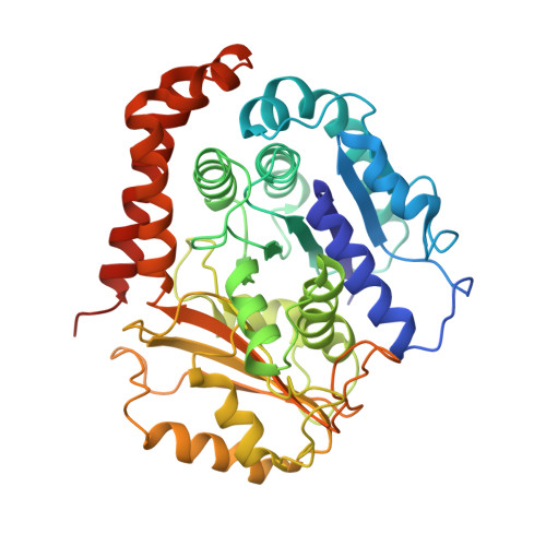
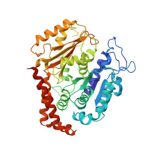
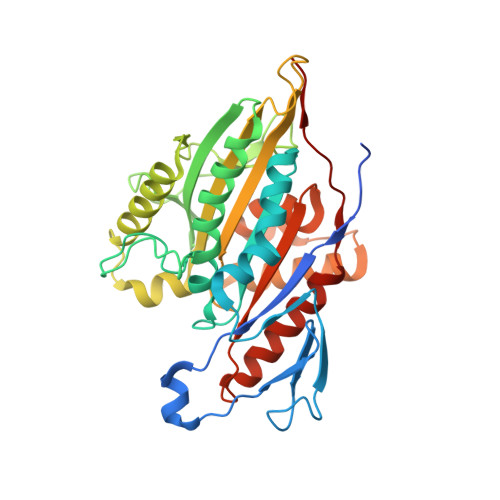
9-Angstrom Structure of a Microtubule-Bound Mitotic Motor.

Bodey, A.J.Kikkawa, M.Moores, C.A.

(2009) J Mol Biology 388: 218

  • DOI: https://doi.org/10.1016/j.jmb.2009.03.008
  • Primary Citation Related Structures: 
    2WBE

  • PubMed Abstract: 

    Kinesin-5 (K5) motors are important components of the microtubule (MT)-based cell division machinery and are targets for small-molecule inhibitors currently in cancer clinical trials. However, the nature of the K5-MT interaction and the regulatory mechanisms that control it remain unclear. Using cryo-electron microscopy and image processing, we calculated the structure of a K5 motor bound to MTs at 9 A resolution, providing insight into this important interaction. Our reconstruction reveals the K5 motor domain in an ATP-like conformation in which MT binding induces the conserved nucleotide-sensing switch I and II loops to form a compact subdomain around the bound nucleotide. Our reconstruction also reveals a novel conformation for the K5-specific drug-binding loop 5, suggesting a possible role for it in switching K5s between force generation and diffusional modes of MT binding. Our data thus shed light on regulation of the interaction between spindle components important for chromosome segregation.


  • Organizational Affiliation
    • School of Crystallography, The Institute of Structural and Molecular Biology, Birkbeck College, Malet Street, London WC1E 7HX, UK.

Macromolecule Content 

  • Total Structure Weight: 144.3 kDa 
  • Atom Count: 9,385 
  • Modeled Residue Count: 1,173 
  • Deposited Residue Count: 1,269 
  • Unique protein chains: 3

Macromolecules

Find similar proteins by:|  3D Structure
Entity ID: 1
MoleculeChains  Sequence LengthOrganismDetailsImage
TUBULIN ALPHA-1D CHAIN451Bos taurusMutation(s): 0 
EC: 3.6.5
UniProt
Find proteins for Q2HJ86 (Bos taurus)
Explore Q2HJ86 
Go to UniProtKB:  Q2HJ86
Entity Groups
Sequence Clusters30% Identity50% Identity70% Identity90% Identity95% Identity100% Identity
UniProt GroupQ2HJ86
Sequence Annotations
Expand
Reference Sequence
Find similar proteins by:|  3D Structure
Entity ID: 2
MoleculeChains  Sequence LengthOrganismDetailsImage
TUBULIN BETA-2B CHAIN445Bos taurusMutation(s): 0 
UniProt
Find proteins for Q6B856 (Bos taurus)
Explore Q6B856 
Go to UniProtKB:  Q6B856
Entity Groups
Sequence Clusters30% Identity50% Identity70% Identity90% Identity95% Identity100% Identity
UniProt GroupQ6B856
Sequence Annotations
Expand
Reference Sequence
Find similar proteins by:|  3D Structure
Entity ID: 3
MoleculeChains  Sequence LengthOrganismDetailsImage
BIPOLAR KINESIN KRP-130373Drosophila melanogasterMutation(s): 0 
UniProt
Find proteins for P46863 (Drosophila melanogaster)
Explore P46863 
Go to UniProtKB:  P46863
Entity Groups
Sequence Clusters30% Identity50% Identity70% Identity90% Identity95% Identity100% Identity
UniProt GroupP46863
Sequence Annotations
Expand
Reference Sequence

Small Molecules

Ligands 5 Unique
IDChains Name / Formula / InChI Key2D Diagram3D Interactions
TA1

Query on TA1


Download:Ideal Coordinates CCD File
G [auth B]TAXOL
C47 H51 N O14
RCINICONZNJXQF-MZXODVADSA-N
GTP

Query on GTP


Download:Ideal Coordinates CCD File
E [auth A]GUANOSINE-5'-TRIPHOSPHATE
C10 H16 N5 O14 P3
XKMLYUALXHKNFT-UUOKFMHZSA-N
ANP

Query on ANP


Download:Ideal Coordinates CCD File
H [auth C]PHOSPHOAMINOPHOSPHONIC ACID-ADENYLATE ESTER
C10 H17 N6 O12 P3
PVKSNHVPLWYQGJ-KQYNXXCUSA-N
GDP

Query on GDP


Download:Ideal Coordinates CCD File
F [auth B]GUANOSINE-5'-DIPHOSPHATE
C10 H15 N5 O11 P2
QGWNDRXFNXRZMB-UUOKFMHZSA-N
MG

Query on MG


Download:Ideal Coordinates CCD File
D [auth A],
I [auth C]
MAGNESIUM ION
Mg
JLVVSXFLKOJNIY-UHFFFAOYSA-N

Experimental Data & Validation

Experimental Data

  • Method: ELECTRON MICROSCOPY
  • Resolution: 9.40 Å
  • Aggregation State: FILAMENT 
  • Reconstruction Method: HELICAL 
EM Software:
TaskSoftware PackageVersion
RECONSTRUCTIONRuby-Helix

Structure Validation

View Full Validation Report


Entry History 

Deposition Data

Revision History  (Full details and data files)

  • Version 1.0: 2009-03-24
    Type: Initial release
  • Version 1.1: 2013-07-24
    Changes: Database references, Derived calculations, Other, Refinement description, Source and taxonomy, Version format compliance
  • Version 1.2: 2016-07-27
    Changes: Atomic model, Database references, Derived calculations, Non-polymer description, Other, Source and taxonomy, Structure summary
  • Version 1.3: 2017-04-19
    Changes: Other
  • Version 1.4: 2018-10-03
    Changes: Author supporting evidence, Data collection
  • Version 1.5: 2024-05-08
    Changes: Data collection, Database references, Derived calculations, Refinement description