2V02 | pdb_00002v02

Recombinant vertebrate calmodulin complexed with Ba


Experimental Data Snapshot

  • Method: X-RAY DIFFRACTION
  • Resolution: 2.20 Å
  • R-Value Free: 
    0.294 (Depositor) 
  • R-Value Work: 
    0.243 (Depositor) 
  • R-Value Observed: 
    0.246 (Depositor) 

Starting Model: experimental
View more details

wwPDB Validation 3D Report Full Report

Validation slider image for 2V02

This is version 1.5 of the entry. See complete history

Literature

A Structural Insight Into Lead Neurotoxicity and Calmodulin Activation by Heavy Metals.

Kursula, P.Majava, V.

(2007) Acta Crystallogr Sect F Struct Biol Cryst Commun 63: 653

  • DOI: https://doi.org/10.1107/S1744309107034525
  • Primary Citation Related Structures: 
    2V01, 2V02

  • PubMed Abstract: 

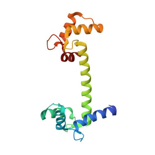
    Calmodulin is a calcium sensor that is also capable of binding and being activated by other metal ions. Of specific interest in this respect is lead, which is known to be neurotoxic and to have a very high affinity towards calmodulin. Crystal structures of human calmodulin complexed with lead and barium ions have been solved. The results will help in understanding the activation mechanisms of calmodulin by different heavy metals and will provide a detailed view of a putative target for lead neurotoxicity in humans.


  • Organizational Affiliation
    • Department of Biochemistry, University of Oulu, Oulu, Finland. petri.kursula@oulu.fi

Macromolecule Content 

  • Total Structure Weight: 17.25 kDa 
  • Atom Count: 1,135 
  • Modeled Residue Count: 142 
  • Deposited Residue Count: 149 
  • Unique protein chains: 1

Macromolecules

Find similar proteins by:|  3D Structure
Entity ID: 1
MoleculeChains  Sequence LengthOrganismDetailsImage
CALMODULIN149Homo sapiensMutation(s): 0 
UniProt & NIH Common Fund Data Resources
Find proteins for P0DP23 (Homo sapiens)
Explore P0DP23 
Go to UniProtKB:  P0DP23
PHAROS:  P0DP23
GTEx:  ENSG00000198668 
Entity Groups
Sequence Clusters30% Identity50% Identity70% Identity90% Identity95% Identity100% Identity
UniProt GroupP0DP23
Sequence Annotations
Expand
Reference Sequence

Experimental Data & Validation

Experimental Data

  • Method: X-RAY DIFFRACTION
  • Resolution: 2.20 Å
  • R-Value Free:  0.294 (Depositor) 
  • R-Value Work:  0.243 (Depositor) 
  • R-Value Observed: 0.246 (Depositor) 
Space Group: P 1
Unit Cell:
Length ( Å )Angle ( ˚ )
a = 24.41α = 89.84
b = 29.32β = 85.68
c = 53.32γ = 82.8
Software Package:
Software NamePurpose
REFMACrefinement
XDSdata reduction
XSCALEdata scaling
MOLREPphasing

Structure Validation

View Full Validation Report


Entry History 

Deposition Data

Revision History  (Full details and data files)

  • Version 1.0: 2007-08-14
    Type: Initial release
  • Version 1.1: 2011-05-08
    Changes: Version format compliance
  • Version 1.2: 2011-07-13
    Changes: Version format compliance
  • Version 1.3: 2018-01-17
    Changes: Data collection
  • Version 1.4: 2018-03-07
    Changes: Source and taxonomy
  • Version 1.5: 2023-12-13
    Changes: Data collection, Database references, Derived calculations, Other, Refinement description