Developmentally Regulated, Combinatorial RNA Processing Modulates Ampa Receptor Biogenesis.
Greger, I.H., Akamine, P., Khatri, L., Ziff, E.B.(2006) Neuron 51: 85
- PubMed: 16815334 Search on PubMed
- DOI: https://doi.org/10.1016/j.neuron.2006.05.020
- Primary Citation Related Structures:
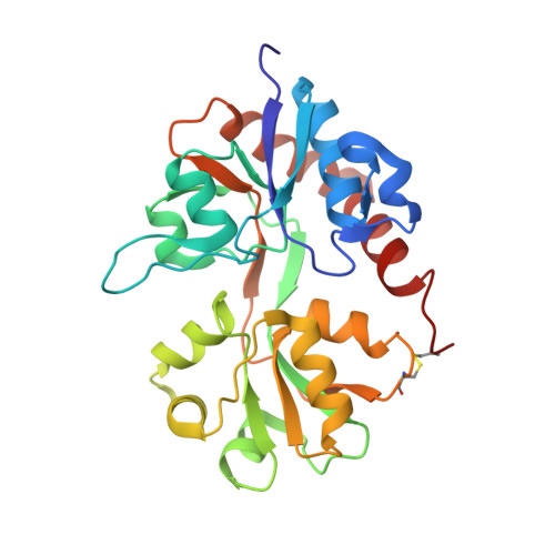
2UXA - PubMed Abstract:
The subunit composition determines AMPA receptor (AMPA-R) function and trafficking. Mechanisms underlying channel assembly are thus central to the efficacy and plasticity of glutamatergic synapses. We previously showed that RNA editing at the Q/R site of the GluR2 subunit contributes to the assembly of AMPA-R heteromers by attenuating formation of GluR2 homotetramers. Here we report that this function of the Q/R site depends on subunit contacts between adjacent ligand binding domains (LBDs). Changes of LBD interface contacts alter GluR2 assembly properties, forward traffic, and expression at synapses. Interestingly, developmentally regulated RNA editing within the LBD (at the R/G site) produces analogous effects. Our data reveal that editing to glycine reduces the self-assembly competence of this critical subunit and slows GluR2 maturation in the endoplasmic reticulum (ER). Therefore, RNA editing sites, located at strategic subunit interfaces, shape AMPA-R assembly and trafficking in a developmentally regulated manner.
- Neurobiology Division, MRC Laboratory of Molecular Biology, Cambridge CB2 2QH, United Kingdom. ig@mrc-lmb.cam.ac.uk
Organizational Affiliation: