


Crystal structure of 3-HSA hydroxylase, oxygenase from Rhodococcus sp. RHA1.
Chang, C., Skarina, T., Kagan, O., Savchenko, A., Edwards, A.M., Joachimiak, A.To be published.
Experimental Data Snapshot
Entity ID: 1 | |||||
|---|---|---|---|---|---|
| Molecule | Chains | Sequence Length | Organism | Details | Image |
| 3-HSA hydroxylase, oxygenase | 394 | Rhodococcus jostii RHA1 | Mutation(s): 0 Gene Names: hsaA, RHA1_ro04539 EC: 1.14.14.12 | 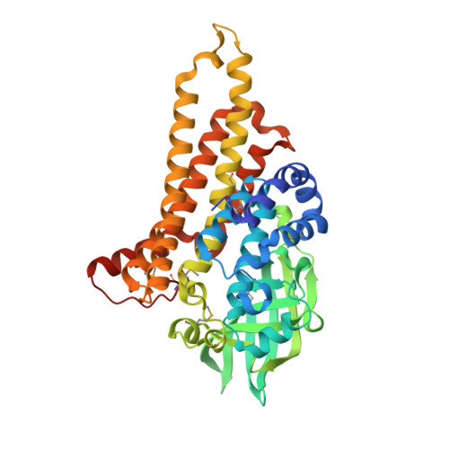 | |
UniProt | |||||
Entity Groups | |||||
| Sequence Clusters | 30% Identity50% Identity70% Identity90% Identity95% Identity100% Identity | ||||
| UniProt Group | Q0S811 | ||||
Sequence AnnotationsExpand | |||||
Reference Sequence | |||||
| Ligands 1 Unique | |||||
|---|---|---|---|---|---|
| ID | Chains | Name / Formula / InChI Key | 2D Diagram | 3D Interactions | |
| 1PS Download:Ideal Coordinates CCD File | E [auth A] F [auth A] G [auth B] H [auth B] I [auth B] | 3-PYRIDINIUM-1-YLPROPANE-1-SULFONATE C8 H11 N O3 S REEBJQTUIJTGAL-UHFFFAOYSA-N |  | ||
| Modified Residues 1 Unique | |||||
|---|---|---|---|---|---|
| ID | Chains | Type | Formula | 2D Diagram | Parent |
| MSE Query on MSE | A, B, C, D | L-PEPTIDE LINKING | C5 H11 N O2 Se |  | MET |
| Length ( Å ) | Angle ( ˚ ) |
|---|---|
| a = 73.283 | α = 83.78 |
| b = 76.137 | β = 75.95 |
| c = 80.881 | γ = 89.99 |
| Software Name | Purpose |
|---|---|
| REFMAC | refinement |
| SBC-Collect | data collection |
| HKL-3000 | data reduction |
| HKL-3000 | data scaling |
| HKL-3000 | phasing |


