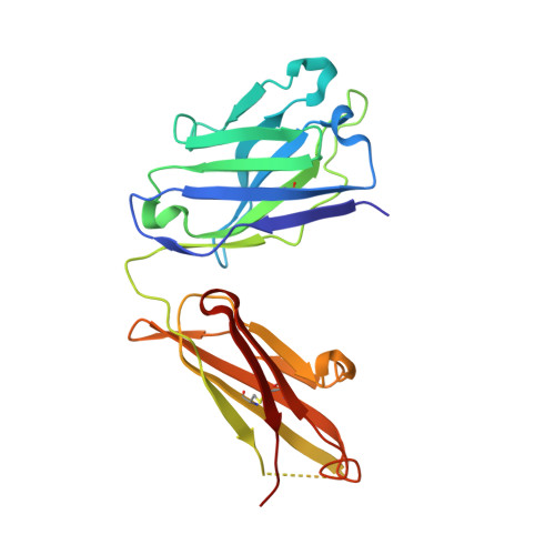
2R2H

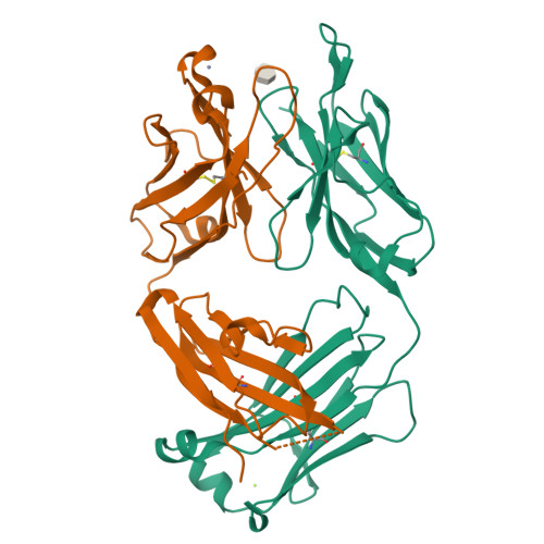
Structure of S25-2 in Complex with Ko


Experimental Data Snapshot

  • Method: X-RAY DIFFRACTION
  • Resolution: 2.00 Å
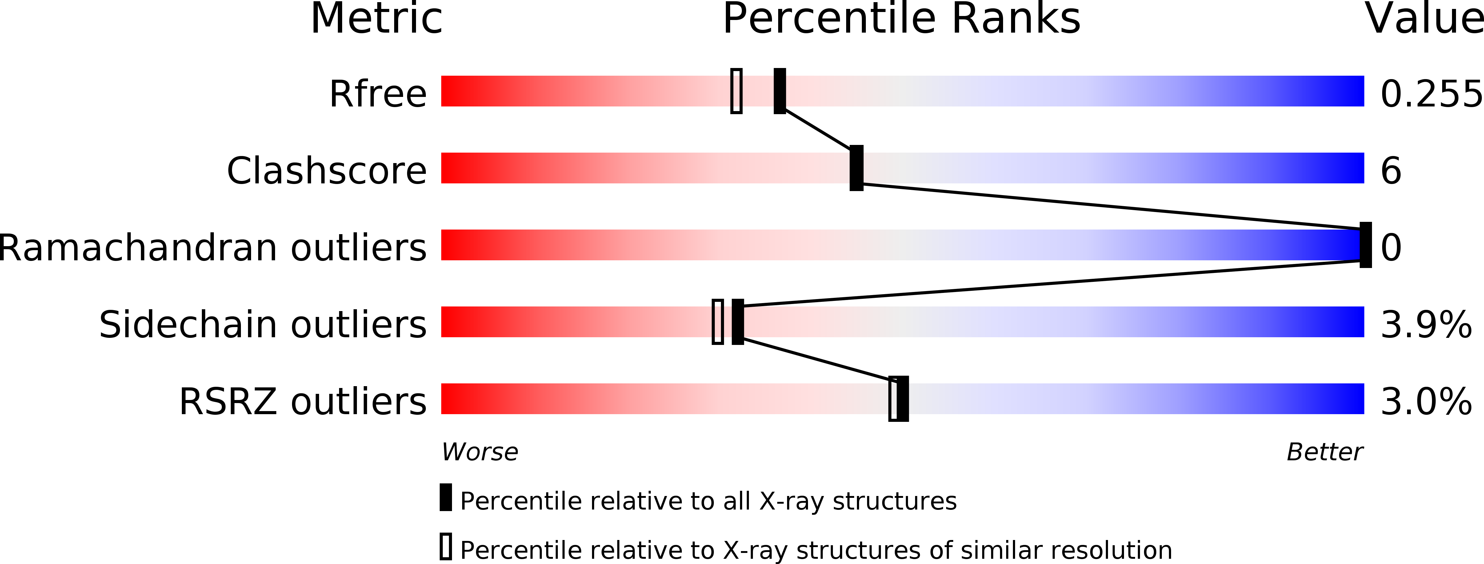
  • R-Value Free: 0.262 
  • R-Value Work: 0.215 
  • R-Value Observed: 0.217 

wwPDB Validation   3D Report Full Report


Ligand Structure Quality Assessment 


This is version 1.4 of the entry. See complete history


Literature

Exploration of specificity in germline monoclonal antibody recognition of a range of natural and synthetic epitopes.

Brooks, C.L.Muller-Loennies, S.Brade, L.Kosma, P.Hirama, T.MacKenzie, C.R.Brade, H.Evans, S.V.

(2008) J Mol Biol 377: 450-468

  • DOI: https://doi.org/10.1016/j.jmb.2008.01.018
  • Primary Citation of Related Structures:  
    2R1W, 2R1X, 2R1Y, 2R23, 2R2B, 2R2E, 2R2H, 3BPC

  • PubMed Abstract: 

    To explore the molecular basis of antigen recognition by germline antibodies, we have determined to high resolution the structures of the near-germline monoclonal antibody S25-2 in complex with seven distinct carbohydrate antigens based on the bacterial sugar 3-deoxy-alpha-D-manno-oct-2-ulosonic acid (Kdo). In contrast to previous findings, the inherited germline Kdo monosaccharide binding site is not restricted to this bacterial sugar but is able to accommodate an array of substitutions and chemical modifications of Kdo, including naturally occurring antigens containing the related monosaccharide d-glycero-alpha-d-talo-oct-2-ulosonic acid as well as nonterminal Kdo residues. However, we show by surface plasmon resonance and ELISA how antibody S25-2 specificity is so dependent on the context in which the antigen is presented that a free disaccharide displays strong binding while the same lipid-A-bound disaccharide does not bind. These structures provide insight into how inherited germline genes code for immunoglobulins of limited flexibility that are capable of binding a range of epitopes from which affinity-matured antibodies are generated.


  • Organizational Affiliation

    Department of Biochemistry and Microbiology, University of Victoria, PO Box 3055 STN CSC, Victoria, BC, Canada.


Macromolecules
Find similar proteins by:  (by identity cutoff)  |  3D Structure
Entity ID: 1
MoleculeChains Sequence LengthOrganismDetailsImage
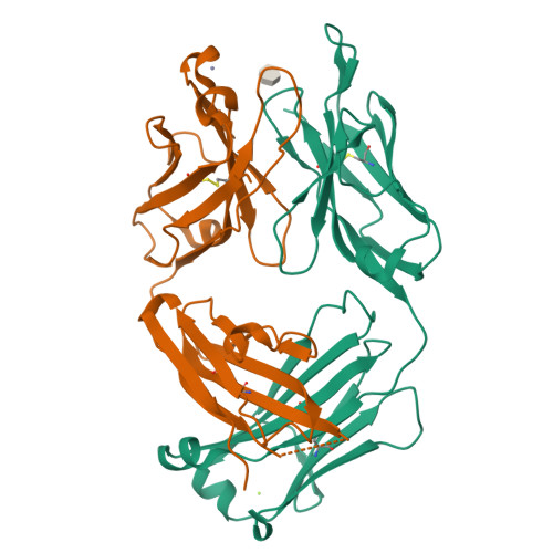
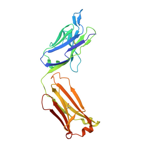
Fab, antibody fragment (IgG1k), light chain219Mus musculusMutation(s): 0 
UniProt
Find proteins for Q52L64 (Mus musculus)
Explore Q52L64 
Go to UniProtKB:  Q52L64
Entity Groups  
Sequence Clusters30% Identity50% Identity70% Identity90% Identity95% Identity100% Identity
UniProt GroupQ52L64
Sequence Annotations
Expand
  • Reference Sequence
Find similar proteins by:  (by identity cutoff)  |  3D Structure
Entity ID: 2
MoleculeChains Sequence LengthOrganismDetailsImage
Fab, antibody fragment (IgG1k), heavy chain222Mus musculusMutation(s): 0 
UniProt
Find proteins for Q6PIP8 (Mus musculus)
Explore Q6PIP8 
Go to UniProtKB:  Q6PIP8
Entity Groups  
Sequence Clusters30% Identity50% Identity70% Identity90% Identity95% Identity100% Identity
UniProt GroupQ6PIP8
Sequence Annotations
Expand
  • Reference Sequence
Experimental Data & Validation

Experimental Data

  • Method: X-RAY DIFFRACTION
  • Resolution: 2.00 Å
  • R-Value Free: 0.262 
  • R-Value Work: 0.215 
  • R-Value Observed: 0.217 
  • Space Group: P 21 21 21
Unit Cell:
Length ( Å )Angle ( ˚ )
a = 45.8α = 90
b = 81.3β = 90
c = 132.35γ = 90
Software Package:
Software NamePurpose
d*TREKdata scaling
PHASERphasing
REFMACrefinement
PDB_EXTRACTdata extraction
d*TREKdata reduction

Structure Validation

View Full Validation Report


Ligand Structure Quality Assessment 


Entry History 

Deposition Data

Revision History  (Full details and data files)

  • Version 1.0: 2008-12-30
    Type: Initial release
  • Version 1.1: 2011-07-13
    Changes: Version format compliance
  • Version 1.2: 2018-03-07
    Changes: Data collection
  • Version 1.3: 2020-07-29
    Type: Remediation
    Reason: Carbohydrate remediation
    Changes: Data collection, Derived calculations
  • Version 1.4: 2024-11-06
    Changes: Data collection, Database references, Structure summary