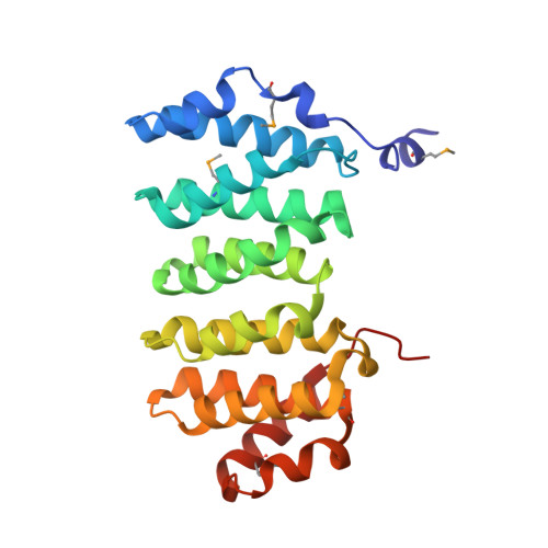
2QK2

Structural Basis of Microtubule Plus End Tracking by XMAP215, CLIP-170 and EB1


Experimental Data Snapshot

  • Method: X-RAY DIFFRACTION
  • Resolution: 2.10 Å
  • R-Value Free: 0.217 
  • R-Value Work: 0.202 

wwPDB Validation   3D Report Full Report


This is version 1.3 of the entry. See complete history


Literature

Structural Basis of Microtubule Plus End Tracking by XMAP215, CLIP-170, and EB1.

Slep, K.C.Vale, R.D.

(2007) Mol Cell 27: 976-991

  • DOI: https://doi.org/10.1016/j.molcel.2007.07.023
  • Primary Citation of Related Structures:  
    2QJX, 2QJZ, 2QK0, 2QK1, 2QK2

  • PubMed Abstract: 

    Microtubule plus end binding proteins (+TIPs) localize to the dynamic plus ends of microtubules, where they stimulate microtubule growth and recruit signaling molecules. Three main +TIP classes have been identified (XMAP215, EB1, and CLIP-170), but whether they act upon microtubule plus ends through a similar mechanism has not been resolved. Here, we report crystal structures of the tubulin binding domains of XMAP215 (yeast Stu2p and Drosophila Msps), EB1 (yeast Bim1p and human EB1), and CLIP-170 (human), which reveal diverse tubulin binding interfaces. Functional studies, however, reveal a common property that native or artificial dimerization of tubulin binding domains (including chemically induced heterodimers of EB1 and CLIP-170) induces tubulin nucleation/assembly in vitro and, in most cases, plus end tracking in living cells. We propose that +TIPs, although diverse in structure, share a common property of multimerizing tubulin, thus acting as polymerization chaperones that aid in subunit addition to the microtubule plus end.


  • Organizational Affiliation

    Howard Hughes Medical Institute, University of California, San Francisco, San Francisco, CA 94158, USA.


Macromolecules
Find similar proteins by:  (by identity cutoff)  |  3D Structure
Entity ID: 1
MoleculeChains Sequence LengthOrganismDetailsImage
LP04448p242Drosophila melanogasterMutation(s): 0 
Gene Names: msps
UniProt
Find proteins for Q9VEZ3 (Drosophila melanogaster)
Explore Q9VEZ3 
Go to UniProtKB:  Q9VEZ3
Entity Groups  
Sequence Clusters30% Identity50% Identity70% Identity90% Identity95% Identity100% Identity
UniProt GroupQ9VEZ3
Sequence Annotations
Expand
  • Reference Sequence
Small Molecules
Modified Residues  1 Unique
IDChains TypeFormula2D DiagramParent
MSE
Query on MSE
A
L-PEPTIDE LINKINGC5 H11 N O2 SeMET
Experimental Data & Validation

Experimental Data

  • Method: X-RAY DIFFRACTION
  • Resolution: 2.10 Å
  • R-Value Free: 0.217 
  • R-Value Work: 0.202 
  • Space Group: C 2 2 21
Unit Cell:
Length ( Å )Angle ( ˚ )
a = 91.37α = 90
b = 113.414β = 90
c = 78.556γ = 90
Software Package:
Software NamePurpose
DENZOdata reduction
SCALEPACKdata scaling
CNSphasing
CNSrefinement
PDB_EXTRACTdata extraction
Blu-Icedata collection

Structure Validation

View Full Validation Report


Entry History 

Deposition Data

Revision History  (Full details and data files)

  • Version 1.0: 2007-10-02
    Type: Initial release
  • Version 1.1: 2011-07-13
    Changes: Version format compliance
  • Version 1.2: 2017-10-18
    Changes: Refinement description
  • Version 1.3: 2024-10-30
    Changes: Data collection, Database references, Derived calculations, Structure summary