2QH7 | pdb_00002qh7

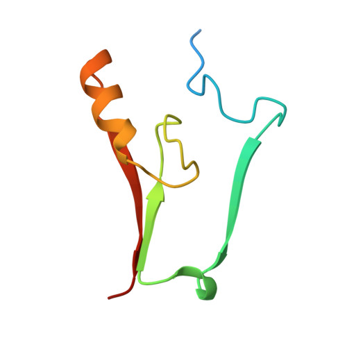
MitoNEET is a uniquely folded 2Fe-2S outer mitochondrial membrane protein stabilized by pioglitazone


Experimental Data Snapshot

  • Method: X-RAY DIFFRACTION
  • Resolution: 1.50 Å
  • R-Value Free: 
    0.223 (Depositor) 
  • R-Value Work: 
    0.182 (Depositor), 0.201 (DCC) 
  • R-Value Observed: 
    0.184 (Depositor) 

wwPDB Validation 3D Report Full Report

Validation slider image for 2QH7

This is version 1.4 of the entry. See complete history. 

Literature

MitoNEET is a uniquely folded 2Fe 2S outer mitochondrial membrane protein stabilized by pioglitazone.

Paddock, M.L., Wiley, S.E., Axelrod, H.L., Cohen, A.E., Roy, M., Abresch, E.C., Capraro, D., Murphy, A.N., Nechushtai, R., Dixon, J.E., Jennings, P.A.

(2007) Proc Natl Acad Sci U S A 104: 14342-14347

  • DOI: https://doi.org/10.1073/pnas.0707189104
  • Primary Citation Related Structures: 
    2QH7

  • PubMed Abstract: 

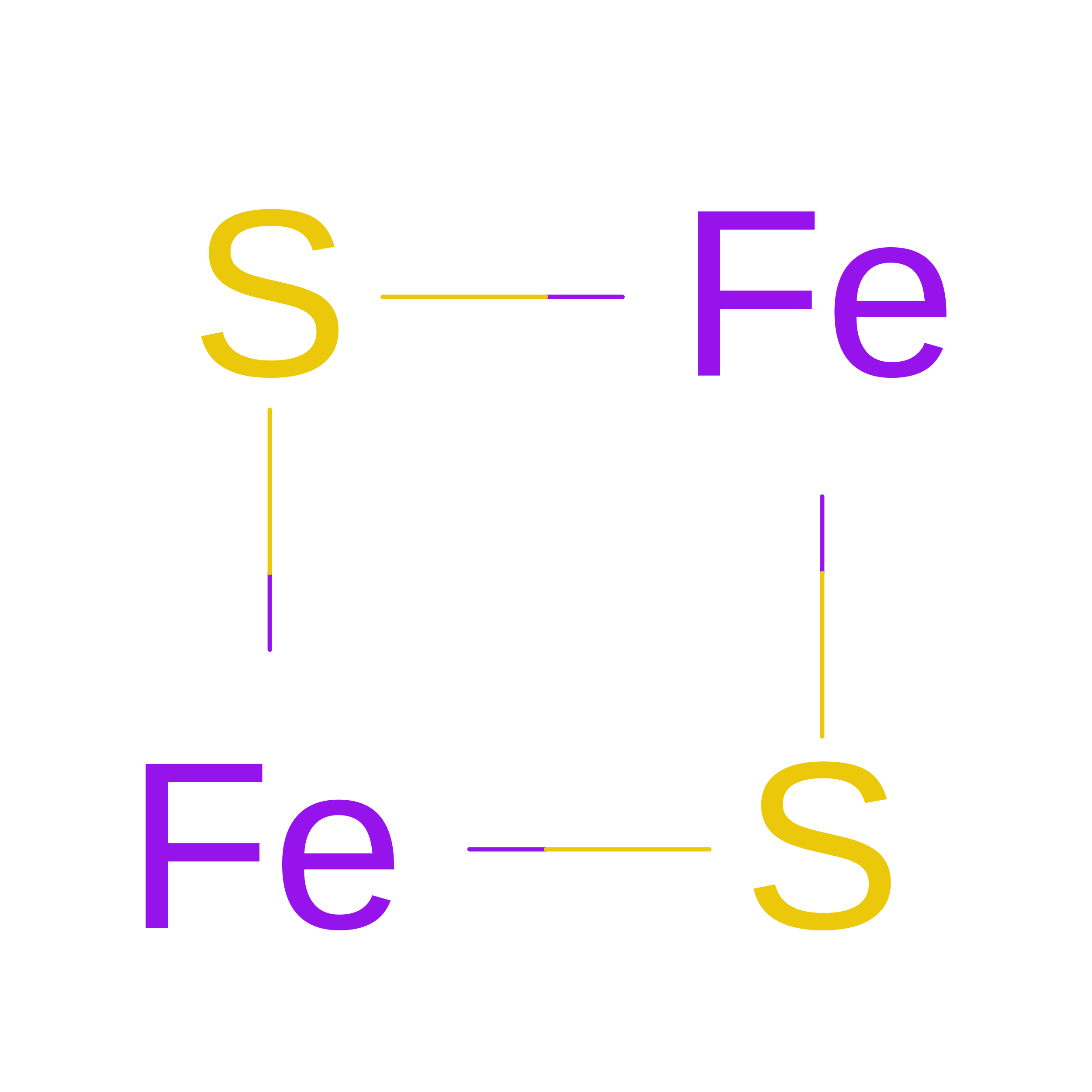
    Iron-sulfur (Fe-S) proteins are key players in vital processes involving energy homeostasis and metabolism from the simplest to most complex organisms. We report a 1.5 A x-ray crystal structure of the first identified outer mitochondrial membrane Fe-S protein, mitoNEET. Two protomers intertwine to form a unique dimeric structure that constitutes a new fold to not only the approximately 650 reported Fe-S protein structures but also to all known proteins. We name this motif the NEET fold. The protomers form a two-domain structure: a beta-cap domain and a cluster-binding domain that coordinates two acid-labile 2Fe-2S clusters. Binding of pioglitazone, an insulin-sensitizing thiazolidinedione used in the treatment of type 2 diabetes, stabilizes the protein against 2Fe-2S cluster release. The biophysical properties of mitoNEET suggest that it may participate in a redox-sensitive signaling and/or in Fe-S cluster transfer.


  • Organizational Affiliation: 
    • Department of Physics, University of California at San Diego, La Jolla, CA 92093, USA.

Macromolecule Content 

  • Total Structure Weight: 18.25 kDa 
  • Atom Count: 1,199 
  • Modeled Residue Count: 130 
  • Deposited Residue Count: 152 
  • Unique protein chains: 1

Macromolecules

Find similar proteins by:|  3D Structure
Entity ID: 1
MoleculeChains  Sequence LengthOrganismDetailsImage
Zinc finger CDGSH-type domain 1
A, B
76Homo sapiensMutation(s): 0 
Gene Names: ZCD1
EC: 2.6.1.3
UniProt & NIH Common Fund Data Resources
Find proteins for Q9NZ45 (Homo sapiens)
Explore Q9NZ45 
Go to UniProtKB:  Q9NZ45
PHAROS:  Q9NZ45
GTEx:  ENSG00000122873 
Entity Groups
Sequence Clusters30% Identity50% Identity70% Identity90% Identity95% Identity100% Identity
UniProt GroupQ9NZ45
Sequence Annotations
Expand
Reference Sequence

Experimental Data & Validation

Experimental Data

  • Method: X-RAY DIFFRACTION
  • Resolution: 1.50 Å
  • R-Value Free:  0.223 (Depositor) 
  • R-Value Work:  0.182 (Depositor), 0.201 (DCC) 
  • R-Value Observed: 0.184 (Depositor) 
Space Group: P 21 21 21
Unit Cell:
Length ( Å )Angle ( ˚ )
a = 46.806α = 90
b = 49.621β = 90
c = 59.006γ = 90
Software Package:
Software NamePurpose
XSCALEdata scaling
SOLVEphasing
RESOLVEphasing
REFMACrefinement
PDB_EXTRACTdata extraction
Blu-Icedata collection
MOSFLMdata reduction
XDSdata reduction
SCALAdata scaling

Structure Validation

View Full Validation Report


Entry History 

Revision History  (Full details and data files)

  • Version 1.0: 2007-08-21
    Type: Initial release
  • Version 1.1: 2011-07-13
    Changes: Advisory, Derived calculations, Version format compliance
  • Version 1.2: 2018-01-24
    Changes: Structure summary
  • Version 1.3: 2024-02-21
    Changes: Data collection, Database references, Derived calculations, Refinement description
  • Version 1.4: 2026-08-12
    Type: Remediation
    Reason: Metalloprotein remediation
    Changes: Derived calculations, Structure summary