2OK2 | pdb_00002ok2

MutS C-terminal domain fused to Maltose Binding Protein


Experimental Data Snapshot

  • Method: X-RAY DIFFRACTION
  • Resolution: 2.00 Å
  • R-Value Free: 
    0.277 (Depositor), 0.277 (DCC) 
  • R-Value Work: 
    0.201 (Depositor), 0.201 (DCC) 
  • R-Value Observed: 
    0.205 (Depositor) 

Starting Model: experimental
View more details

wwPDB Validation 3D Report Full Report

Validation slider image for 2OK2

This is version 2.1 of the entry. See complete history

Literature

Escherichia coli MutS Tetramerization Domain Structure Reveals That Stable Dimers but Not Tetramers Are Essential for DNA Mismatch Repair in Vivo.

Mendillo, M.L.Putnam, C.D.Kolodner, R.D.

(2007) J Biological Chem 282: 16345-16354

  • DOI: https://doi.org/10.1074/jbc.M700858200
  • Primary Citation Related Structures: 
    2OK2

  • PubMed Abstract: 

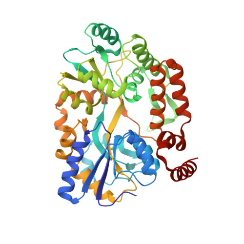
    The Escherichia coli mispair-binding protein MutS forms dimers and tetramers in vitro, although the functional form in vivo is under debate. Here we demonstrate that the MutS tetramer is extended in solution using small angle x-ray scattering and the crystal structure of the C-terminal 34 amino acids of MutS containing the tetramer-forming domain fused to maltose-binding protein (MBP). Wild-type C-terminal MBP fusions formed tetramers and could bind MutS and MutS-MutL-DNA complexes. In contrast, D835R and R840E mutations predicted to disrupt tetrameric interactions only allowed dimerization of MBP. A chromosomal MutS truncation mutation eliminating the dimerization/tetramerization domain eliminated mismatch repair, whereas the tetramer-disrupting MutS D835R and R840E mutations only modestly affected MutS function. These results demonstrate that dimerization but not tetramerization of the MutS C terminus is essential for mismatch repair.


  • Organizational Affiliation
    • Ludwig Institute for Cancer Research, Department of Medicine, University of California, San Diego School of Medicine, La Jolla, California 92093-0669, USA.

Macromolecule Content 

  • Total Structure Weight: 89.57 kDa 
  • Atom Count: 6,889 
  • Modeled Residue Count: 804 
  • Deposited Residue Count: 804 
  • Unique protein chains: 1

Macromolecules

Find similar proteins by:|  3D Structure
Entity ID: 1
MoleculeChains  Sequence LengthOrganismDetailsImage
Maltose-binding periplasmic protein, DNA mismatch repair protein mutS fusion protein
A, B
402Escherichia coli K-12Mutation(s): 0 
Gene Names: malE
UniProt
Find proteins for P23909 (Escherichia coli (strain K12))
Explore P23909 
Go to UniProtKB:  P23909
Entity Groups
Sequence Clusters30% Identity50% Identity70% Identity90% Identity95% Identity100% Identity
UniProt GroupP23909
Sequence Annotations
Expand
Reference Sequence

Oligosaccharides

Help  
Entity ID: 2
MoleculeChains Length2D Diagram GlycosylationD Interactions
alpha-D-glucopyranose-(1-4)-alpha-D-glucopyranose
C, D
2N/A
Glycosylation Resources
GlyTouCan: G07411ON
GlyCosmos: G07411ON

Small Molecules

Ligands 1 Unique
IDChains Name / Formula / InChI Key2D Diagram3D Interactions
SO4

Query on SO4


Download:Ideal Coordinates CCD File
E [auth A]SULFATE ION
O4 S
QAOWNCQODCNURD-UHFFFAOYSA-L

Biologically Interesting Molecules (External Reference) 

1 Unique

Experimental Data & Validation

Experimental Data

  • Method: X-RAY DIFFRACTION
  • Resolution: 2.00 Å
  • R-Value Free:  0.277 (Depositor), 0.277 (DCC) 
  • R-Value Work:  0.201 (Depositor), 0.201 (DCC) 
  • R-Value Observed: 0.205 (Depositor) 
Space Group: C 1 2 1
Unit Cell:
Length ( Å )Angle ( ˚ )
a = 174.289α = 90
b = 88.361β = 106.8
c = 61.353γ = 90
Software Package:
Software NamePurpose
REFMACrefinement
Blu-Icedata collection
DENZOdata reduction
SCALEPACKdata scaling
AMoREphasing
XFITdata reduction

Structure Validation

View Full Validation Report


Entry History 

Deposition Data

Revision History  (Full details and data files)

  • Version 1.0: 2007-04-10
    Type: Initial release
  • Version 1.1: 2008-05-01
    Changes: Version format compliance
  • Version 1.2: 2011-07-13
    Changes: Advisory, Version format compliance
  • Version 1.3: 2017-08-23
    Changes: Refinement description, Source and taxonomy
  • Version 2.0: 2020-07-29
    Type: Remediation
    Reason: Carbohydrate remediation
    Changes: Atomic model, Data collection, Database references, Derived calculations, Non-polymer description, Structure summary
  • Version 2.1: 2023-08-30
    Changes: Data collection, Database references, Refinement description, Structure summary