2O17 | pdb_00002o17

Pectate lyase bound to hexasaccharide


Experimental Data Snapshot

  • Method: X-RAY DIFFRACTION
  • Resolution: 2.30 Å
  • R-Value Free: 
    0.223 (Depositor), 0.223 (DCC) 
  • R-Value Work: 
    0.143 (Depositor), 0.144 (DCC) 
  • R-Value Observed: 
    0.147 (Depositor) 

wwPDB Validation 3D Report Full Report

Validation slider image for 2O17

This is version 2.2 of the entry. See complete history

Literature

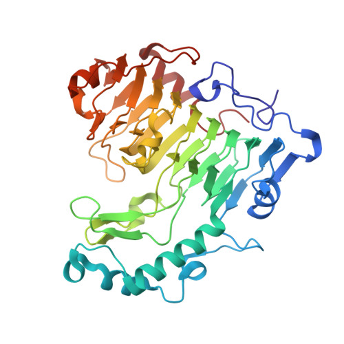
Structural insights into substrate specificity and the anti beta-elimination mechanism of pectate lyase.

Seyedarabi, A.To, T.T.Ali, S.Hussain, S.Fries, M.Madsen, R.Clausen, M.H.Teixteira, S.Brocklehurst, K.Pickersgill, R.W.

(2010) Biochemistry 49: 539-546

  • DOI: https://doi.org/10.1021/bi901503g
  • Primary Citation Related Structures: 
    2NZM, 2O04, 2O0V, 2O17, 2O1D, 3KRG

  • PubMed Abstract: 

    Pectate lyases harness anti beta-elimination chemistry to cleave the alpha-1,4 linkage in the homogalacturonan region of plant cell wall pectin. We have studied the binding of five pectic oligosaccharides to Bacillus subtilis pectate lyase in crystals of the inactive enzyme in which the catalytic base is substituted with alanine (R279A). We discover that the three central subsites (-1, +1, and +2) have a profound preference for galacturonate but that the distal subsites can accommodate methylated galacturonate. It is reasonable to assume therefore that pectate lyase can cleave pectin with three consecutive galacturonate residues. The enzyme in the absence of substrate binds a single calcium ion, and we show that two additional calcium ions bind between enzyme and substrate carboxylates occupying the +1 subsite in the Michaelis complex. The substrate binds less intimately to the enzyme in a complex made with a catalytic base in place but in the absence of the calcium ions and an adjacent lysine. In this complex, the catalytic base is correctly positioned to abstract the C5 proton, but there are no calcium ions binding the carboxylate at the +1 subsite. It is clear, therefore, that the catalytic calcium ions and adjacent lysine promote catalysis by acidifying the alpha-proton, facilitating its abstraction by the base. There is also clear evidence that binding distorts the relaxed 2(1) or 3(1) helical conformation of the oligosaccharides in the region of the scissile bond.


  • Organizational Affiliation
    • School of Biological and Chemical Sciences, Queen Mary University of London, Mile End Road, London E1 4NS, United Kingdom.

Macromolecule Content 

  • Total Structure Weight: 44.38 kDa 
  • Atom Count: 3,640 
  • Modeled Residue Count: 399 
  • Deposited Residue Count: 399 
  • Unique protein chains: 1

Macromolecules

Find similar proteins by:|  3D Structure
Entity ID: 1
MoleculeChains  Sequence LengthOrganismDetailsImage
Pectate lyase399Bacillus subtilisMutation(s): 1 
Gene Names: pel
EC: 4.2.2.2
UniProt
Find proteins for P39116 (Bacillus subtilis (strain 168))
Explore P39116 
Go to UniProtKB:  P39116
Entity Groups
Sequence Clusters30% Identity50% Identity70% Identity90% Identity95% Identity100% Identity
UniProt GroupP39116
Sequence Annotations
Expand
Reference Sequence

Oligosaccharides

Help  
Entity ID: 2
MoleculeChains Length2D Diagram GlycosylationD Interactions
alpha-D-galactopyranuronic acid-(1-4)-alpha-D-galactopyranuronic acid-(1-4)-alpha-D-galactopyranuronic acid-(1-4)-alpha-D-galactopyranuronic acid-(1-4)-alpha-D-galactopyranuronic acid
B
5N/A
Glycosylation Resources
GlyTouCan: G48720UK
GlyCosmos: G48720UK

Experimental Data & Validation

Experimental Data

  • Method: X-RAY DIFFRACTION
  • Resolution: 2.30 Å
  • R-Value Free:  0.223 (Depositor), 0.223 (DCC) 
  • R-Value Work:  0.143 (Depositor), 0.144 (DCC) 
  • R-Value Observed: 0.147 (Depositor) 
Space Group: P 1 21 1
Unit Cell:
Length ( Å )Angle ( ˚ )
a = 51.254α = 90
b = 69.345β = 113.69
c = 59.826γ = 90
Software Package:
Software NamePurpose
REFMACrefinement
MOSFLMdata reduction
SCALAdata scaling
MOLREPphasing

Structure Validation

View Full Validation Report


Entry History 

Deposition Data

Revision History  (Full details and data files)

  • Version 1.0: 2007-11-20
    Type: Initial release
  • Version 1.1: 2011-07-13
    Changes: Version format compliance
  • Version 2.0: 2020-07-29
    Type: Remediation
    Reason: Carbohydrate remediation
    Changes: Atomic model, Data collection, Derived calculations, Structure summary
  • Version 2.1: 2021-10-20
    Changes: Database references, Structure summary
  • Version 2.2: 2023-12-27
    Changes: Data collection