2JH1 | pdb_00002jh1

Crystal structure of Toxoplasma gondii micronemal protein 1


Experimental Data Snapshot

  • Method: X-RAY DIFFRACTION
  • Resolution: 1.90 Å
  • R-Value Free: 
    0.205 (Depositor), 0.204 (DCC) 
  • R-Value Work: 
    0.178 (Depositor), 0.176 (DCC) 
  • R-Value Observed: 
    0.179 (Depositor) 

wwPDB Validation 3D Report Full Report

Validation slider image for 2JH1

This is version 1.4 of the entry. See complete history

Literature

Atomic Resolution Insight Into Host Cell Recognition by Toxoplasma Gondii.

Blumenschein, T.M.A.Friedrich, N.Childs, R.A.Saouros, S.Carpenter, E.P.Campanero-Rhodes, M.A.Simpson, P.Chai, W.Koutroukides, T.Blackman, M.J.Feizi, T.Soldati-Favre, D.Matthews, S.

(2007) EMBO J 26: 2808

  • DOI: https://doi.org/10.1038/sj.emboj.7601704
  • Primary Citation Related Structures: 
    2JH1, 2JH7, 2JHD

  • PubMed Abstract: 

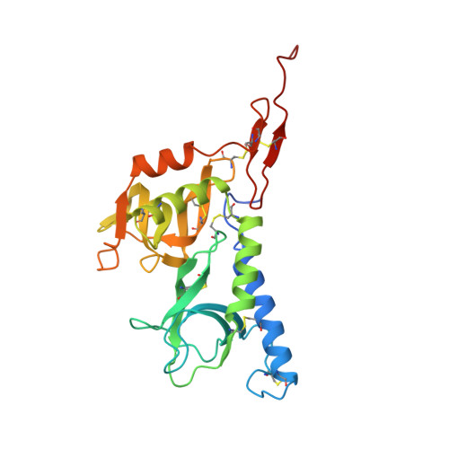
    The obligate intracellular parasite Toxoplasma gondii, a member of the phylum Apicomplexa that includes Plasmodium spp., is one of the most widespread parasites and the causative agent of toxoplasmosis. Micronemal proteins (MICs) are released onto the parasite surface just before invasion of host cells and play important roles in host cell recognition, attachment and penetration. Here, we report the atomic structure for a key MIC, TgMIC1, and reveal a novel cell-binding motif called the microneme adhesive repeat (MAR). Using glycoarray analyses, we identified a novel interaction with sialylated oligosaccharides that resolves several prevailing misconceptions concerning TgMIC1. Structural studies of various complexes between TgMIC1 and sialylated oligosaccharides provide high-resolution insights into the recognition of sialylated oligosaccharides by a parasite surface protein. We observe that MAR domains exist in tandem repeats, which provide a highly specialized structure for glycan discrimination. Our work uncovers new features of parasite-receptor interactions at the early stages of host cell invasion, which will assist the design of new therapeutic strategies.


  • Organizational Affiliation
    • Division of Molecular Biosciences, Imperial College London, London, UK.

Macromolecule Content 

  • Total Structure Weight: 28.31 kDa 
  • Atom Count: 2,094 
  • Modeled Residue Count: 235 
  • Deposited Residue Count: 246 
  • Unique protein chains: 1

Macromolecules

Find similar proteins by:|  3D Structure
Entity ID: 1
MoleculeChains  Sequence LengthOrganismDetailsImage
MICRONEMAL PROTEIN 1246Toxoplasma gondiiMutation(s): 0 
UniProt
Find proteins for O00834 (Toxoplasma gondii)
Explore O00834 
Go to UniProtKB:  O00834
Entity Groups
Sequence Clusters30% Identity50% Identity70% Identity90% Identity95% Identity100% Identity
UniProt GroupO00834
Sequence Annotations
Expand
Reference Sequence

Small Molecules

Ligands 3 Unique
IDChains Name / Formula / InChI Key2D Diagram3D Interactions
GOL

Query on GOL


Download:Ideal Coordinates CCD File
O [auth A],
T [auth A]
GLYCEROL
C3 H8 O3
PEDCQBHIVMGVHV-UHFFFAOYSA-N
ACT

Query on ACT


Download:Ideal Coordinates CCD File
B [auth A]
C [auth A]
D [auth A]
E [auth A]
F [auth A]
B [auth A],
C [auth A],
D [auth A],
E [auth A],
F [auth A],
G [auth A],
H [auth A],
I [auth A],
J [auth A],
K [auth A],
L [auth A],
P [auth A],
Q [auth A],
R [auth A],
S [auth A]
ACETATE ION
C2 H3 O2
QTBSBXVTEAMEQO-UHFFFAOYSA-M
CL

Query on CL


Download:Ideal Coordinates CCD File
M [auth A],
N [auth A]
CHLORIDE ION
Cl
VEXZGXHMUGYJMC-UHFFFAOYSA-M

Experimental Data & Validation

Experimental Data

  • Method: X-RAY DIFFRACTION
  • Resolution: 1.90 Å
  • R-Value Free:  0.205 (Depositor), 0.204 (DCC) 
  • R-Value Work:  0.178 (Depositor), 0.176 (DCC) 
  • R-Value Observed: 0.179 (Depositor) 
Space Group: P 43 21 2
Unit Cell:
Length ( Å )Angle ( ˚ )
a = 66.192α = 90
b = 66.192β = 90
c = 172.299γ = 90
Software Package:
Software NamePurpose
REFMACrefinement
DENZOdata reduction
SCALEPACKdata scaling
SOLVEphasing

Structure Validation

View Full Validation Report


Entry History 

Revision History  (Full details and data files)

  • Version 1.0: 2007-05-22
    Type: Initial release
  • Version 1.1: 2011-05-08
    Changes: Version format compliance
  • Version 1.2: 2011-07-13
    Changes: Version format compliance
  • Version 1.3: 2019-01-30
    Changes: Data collection, Derived calculations, Experimental preparation, Other
  • Version 1.4: 2024-10-16
    Changes: Data collection, Database references, Derived calculations, Other, Structure summary