2J0A | pdb_00002j0a

Structure of the catalytic domain of mouse Manic Fringe


Experimental Data Snapshot

  • Method: X-RAY DIFFRACTION
  • Resolution: 1.80 Å
  • R-Value Free: 
    0.219 (Depositor), 0.222 (DCC) 
  • R-Value Work: 
    0.194 (Depositor), 0.197 (DCC) 
  • R-Value Observed: 
    0.195 (Depositor) 

wwPDB Validation 3D Report Full Report

Validation slider image for 2J0A

This is version 1.2 of the entry. See complete history

Literature

Structural Insights Into the Notch-Modifying Glycosyltransferase Fringe

Jinek, M.Chen, Y.-W.Clausen, H.Cohen, S.M.Conti, E.

(2006) Nat Struct Mol Biol 13: 945

  • DOI: https://doi.org/10.1038/nsmb1144
  • Primary Citation Related Structures: 
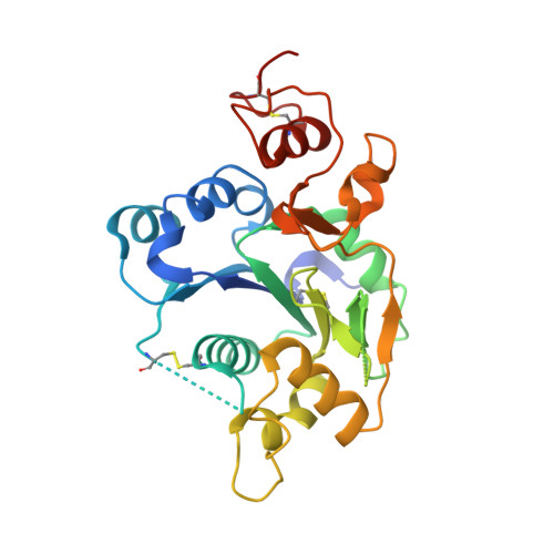
    2J0A, 2J0B

  • PubMed Abstract: 

    Fringe proteins are beta1,3-N-acetylglucosaminyltransferases that modify Notch receptors, altering their ligand-binding specificity to regulate Notch signaling in development. We present the crystal structure of mouse Manic Fringe bound to UDP and manganese. The structure reveals amino acid residues involved in recognition of donor substrates and catalysis, and a putative binding pocket for acceptor substrates. Mutations of several invariant residues in this pocket impair Fringe activity in vivo.


  • Organizational Affiliation
    • European Molecular Biology Laboratory, Meyerhofstrasse 1, D-69117 Heidelberg, Germany.

Macromolecule Content 

  • Total Structure Weight: 31.68 kDa 
  • Atom Count: 2,048 
  • Modeled Residue Count: 243 
  • Deposited Residue Count: 280 
  • Unique protein chains: 1

Macromolecules

Find similar proteins by:|  3D Structure
Entity ID: 1
MoleculeChains  Sequence LengthOrganismDetailsImage
BETA-1,3-N-ACETYLGLUCOSAMINYLTRANSFERASE MANIC FRINGE280Mus musculusMutation(s): 1 
EC: 2.4.1.222
UniProt & NIH Common Fund Data Resources
Find proteins for O09008 (Mus musculus)
Explore O09008 
Go to UniProtKB:  O09008
IMPC:  MGI:1095404
Entity Groups
Sequence Clusters30% Identity50% Identity70% Identity90% Identity95% Identity100% Identity
UniProt GroupO09008
Sequence Annotations
Expand
Reference Sequence

Experimental Data & Validation

Experimental Data

  • Method: X-RAY DIFFRACTION
  • Resolution: 1.80 Å
  • R-Value Free:  0.219 (Depositor), 0.222 (DCC) 
  • R-Value Work:  0.194 (Depositor), 0.197 (DCC) 
  • R-Value Observed: 0.195 (Depositor) 
Space Group: P 21 21 2
Unit Cell:
Length ( Å )Angle ( ˚ )
a = 162.6α = 90
b = 41.4β = 90
c = 39.8γ = 90
Software Package:
Software NamePurpose
REFMACrefinement
XDSdata reduction
XDSdata scaling
PHENIXphasing
HYSSphasing
autoSHARPphasing

Structure Validation

View Full Validation Report


Entry History 

Deposition Data

Revision History  (Full details and data files)

  • Version 1.0: 2006-09-04
    Type: Initial release
  • Version 1.1: 2011-07-13
    Changes: Advisory, Version format compliance
  • Version 1.2: 2024-11-20
    Changes: Data collection, Database references, Derived calculations, Other, Structure summary