2IW4 | pdb_00002iw4

CRYSTAL STRUCTURE OF BASILLUS SUBTILIS FAMILY II INORGANIC PYROPHOSPHATASE MUTANT, H98Q, IN COMPLEX WITH PNP


Experimental Data Snapshot

  • Method: X-RAY DIFFRACTION
  • Resolution: 2.15 Å
  • R-Value Free: 
    0.205 (Depositor), 0.206 (DCC) 
  • R-Value Work: 
    0.169 (Depositor), 0.171 (DCC) 
  • R-Value Observed: 
    0.170 (Depositor) 

Starting Model: experimental
View more details

wwPDB Validation 3D Report Full Report

Validation slider image for 2IW4

Ligand Structure Quality Assessment 


This is version 1.3 of the entry. See complete history

Literature

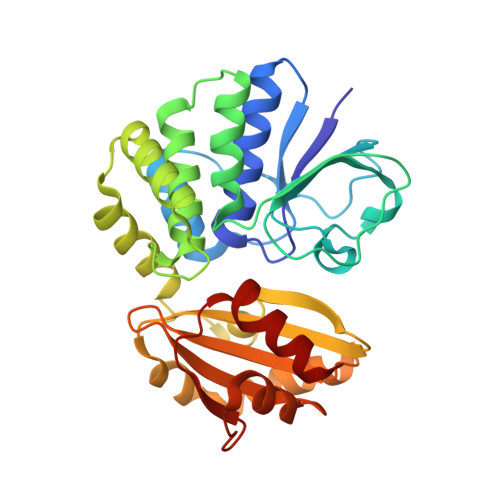
A Trimetal Site and Substrate Distortion in a Family II Inorganic Pyrophosphatase.

Fabrichniy, I.P.Lehtio, L.Tammenkoski, M.Zyryanov, A.B.Oksanen, E.Baykov, A.A.Lahti, R.Goldman, A.

(2007) J Biological Chem 282: 1422

  • DOI: https://doi.org/10.1074/jbc.M513161200
  • Primary Citation Related Structures: 
    2HAW, 2IW4

  • PubMed Abstract: 

    We report the first crystal structures of a family II pyrophosphatase complexed with a substrate analogue, imidodiphosphate (PNP). These provide new insights into the catalytic reaction mechanism of this enzyme family. We were able to capture the substrate complex both by fluoride inhibition and by site-directed mutagenesis providing complementary snapshots of the Michaelis complex. Structures of both the fluoride-inhibited wild type and the H98Q variant of the PNP-Bacillus subtilis pyrophosphatase complex show a unique trinuclear metal center. Each metal ion coordinates a terminal oxygen on the electrophilic phosphate and a lone pair on the putative nucleophile, thus placing it in line with the scissile bond without any coordination by protein. The nucleophile moves further away from the electrophilic phosphorus site, to the opposite side of the trimetal plane, upon binding of substrate. In comparison with earlier product complexes, the side chain of Lys296 has swung in and so three positively charged side chains, His98, Lys205 and Lys296, now surround the bridging nitrogen in PNP. Finally, one of the active sites in the wild-type structure appears to show evidence of substrate distortion. Binding to the enzyme may thus strain the substrate and thus enhance the catalytic rate.


  • Organizational Affiliation
    • Program in Structural Biology and Biophysics, Institute of Biotechnology, University of Helsinki, Biocenter 3, P. O. Box 65, FIN-00014 Helsinki, Finland.

Macromolecule Content 

  • Total Structure Weight: 69.64 kDa 
  • Atom Count: 5,334 
  • Modeled Residue Count: 617 
  • Deposited Residue Count: 618 
  • Unique protein chains: 1

Macromolecules

Find similar proteins by:|  3D Structure
Entity ID: 1
MoleculeChains  Sequence LengthOrganismDetailsImage
MANGANESE-DEPENDENT INORGANIC PYROPHOSPHATASE
A, B
309Bacillus subtilisMutation(s): 1 
EC: 3.6.1.1
UniProt
Find proteins for P37487 (Bacillus subtilis (strain 168))
Explore P37487 
Go to UniProtKB:  P37487
Entity Groups
Sequence Clusters30% Identity50% Identity70% Identity90% Identity95% Identity100% Identity
UniProt GroupP37487
Sequence Annotations
Expand
Reference Sequence

Small Molecules

Ligands 7 Unique
IDChains Name / Formula / InChI Key2D Diagram3D Interactions
PG4

Query on PG4


Download:Ideal Coordinates CCD File
K [auth B],
L [auth B]
TETRAETHYLENE GLYCOL
C8 H18 O5
UWHCKJMYHZGTIT-UHFFFAOYSA-N
2PN

Query on 2PN


Download:Ideal Coordinates CCD File
I [auth A],
T [auth B]
IMIDODIPHOSPHORIC ACID
H5 N O6 P2
GNGSOPFGGKKDQP-UHFFFAOYSA-N
SO4

Query on SO4


Download:Ideal Coordinates CCD File
J [auth A],
U [auth B]
SULFATE ION
O4 S
QAOWNCQODCNURD-UHFFFAOYSA-L
GOL

Query on GOL


Download:Ideal Coordinates CCD File
Q [auth B],
R [auth B]
GLYCEROL
C3 H8 O3
PEDCQBHIVMGVHV-UHFFFAOYSA-N
FE

Query on FE


Download:Ideal Coordinates CCD File
C [auth A],
D [auth A],
M [auth B],
N [auth B]
FE (III) ION
Fe
VTLYFUHAOXGGBS-UHFFFAOYSA-N
MN

Query on MN


Download:Ideal Coordinates CCD File
G [auth A],
H [auth A],
S [auth B]
MANGANESE (II) ION
Mn
WAEMQWOKJMHJLA-UHFFFAOYSA-N
MG

Query on MG


Download:Ideal Coordinates CCD File
E [auth A],
F [auth A],
O [auth B],
P [auth B]
MAGNESIUM ION
Mg
JLVVSXFLKOJNIY-UHFFFAOYSA-N

Experimental Data & Validation

Experimental Data

  • Method: X-RAY DIFFRACTION
  • Resolution: 2.15 Å
  • R-Value Free:  0.205 (Depositor), 0.206 (DCC) 
  • R-Value Work:  0.169 (Depositor), 0.171 (DCC) 
  • R-Value Observed: 0.170 (Depositor) 
Space Group: P 21 21 21
Unit Cell:
Length ( Å )Angle ( ˚ )
a = 59.908α = 90
b = 115.249β = 90
c = 148.06γ = 90
Software Package:
Software NamePurpose
REFMACrefinement
DENZOdata reduction
SCALEPACKdata scaling
CNSphasing

Structure Validation

View Full Validation Report


Ligand Structure Quality Assessment 


Entry History 

Deposition Data

Revision History  (Full details and data files)

  • Version 1.0: 2006-11-07
    Type: Initial release
  • Version 1.1: 2011-07-13
    Changes: Advisory, Version format compliance
  • Version 1.2: 2018-01-24
    Changes: Source and taxonomy
  • Version 1.3: 2023-12-13
    Changes: Data collection, Database references, Derived calculations, Other, Refinement description