2IHO | pdb_00002iho

Crystal structure of MOA, a lectin from the mushroom Marasmius oreades in complex with the trisaccharide Gal(1,3)Gal(1,4)GlcNAc


Experimental Data Snapshot

  • Method: X-RAY DIFFRACTION
  • Resolution: 2.41 Å
  • R-Value Free: 
    0.211 (Depositor), 0.205 (DCC) 
  • R-Value Work: 
    0.190 (Depositor), 0.187 (DCC) 
  • R-Value Observed: 
    0.192 (Depositor) 

wwPDB Validation 3D Report Full Report

Validation slider image for 2IHO

This is version 2.1 of the entry. See complete history

Literature

Crystal structure of the marasmius oreades mushroom lectin in complex with a xenotransplantation epitope.

Grahn, E.Askarieh, G.Holmner, A.Tateno, H.Winter, H.C.Goldstein, I.J.Krengel, U.

(2007) J Mol Biology 369: 710-721

  • DOI: https://doi.org/10.1016/j.jmb.2007.03.016
  • Primary Citation Related Structures: 
    2IHO

  • PubMed Abstract: 

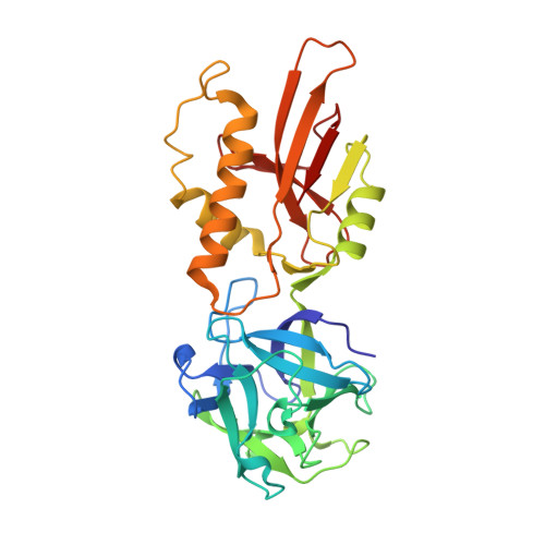
    MOA, a lectin from the mushroom Marasmius oreades, is one of the few reagents that specifically agglutinate blood group B erythrocytes. Further, it is the only lectin known to have exclusive specificity for Galalpha(1,3)Gal-containing sugar epitopes, which are antigens that pose a severe barrier to animal-to-human organ transplantation. We describe here the structure of MOA at 2.4 A resolution, in complex with the linear trisaccharide Galalpha(1,3)Galbeta(1,4)GlcNAc. The structure is dimeric, with two distinct domains per protomer: the N-terminal lectin module adopts a ricinB/beta-trefoil fold and contains three putative carbohydrate-binding sites, while the C-terminal domain serves as a dimerization interface. This latter domain, which has an unknown function, reveals a novel fold with intriguing conservation of an active site cleft. A number of indications suggest that MOA may have an enzymatic function in addition to the sugar-binding properties.


  • Organizational Affiliation
    • Department of Chemistry, University of Oslo, P.O. Box 1033, Blindern, NO-0315 Oslo, Norway. elin.grahn@kjemi.uio.no <elin.grahn@kjemi.uio.no>

Macromolecule Content 

  • Total Structure Weight: 33.42 kDa 
  • Atom Count: 2,563 
  • Modeled Residue Count: 292 
  • Deposited Residue Count: 293 
  • Unique protein chains: 1

Macromolecules

Find similar proteins by:|  3D Structure
Entity ID: 1
MoleculeChains  Sequence LengthOrganismDetailsImage
Lectin293Marasmius oreadesMutation(s): 0 
UniProt
Find proteins for Q8X123 (Marasmius oreades)
Explore Q8X123 
Go to UniProtKB:  Q8X123
Entity Groups
Sequence Clusters30% Identity50% Identity70% Identity90% Identity95% Identity100% Identity
UniProt GroupQ8X123
Sequence Annotations
Expand
Reference Sequence

Oligosaccharides

Help  
Entity ID: 2
MoleculeChains Length2D Diagram GlycosylationD Interactions
beta-D-galactopyranose-(1-3)-beta-D-galactopyranose-(1-4)-2-acetamido-2-deoxy-beta-D-glucopyranose
B, C
3N/A
Glycosylation Resources
GlyTouCan: G35116BF
GlyCosmos: G35116BF
GlyGen: G35116BF

Experimental Data & Validation

Experimental Data

  • Method: X-RAY DIFFRACTION
  • Resolution: 2.41 Å
  • R-Value Free:  0.211 (Depositor), 0.205 (DCC) 
  • R-Value Work:  0.190 (Depositor), 0.187 (DCC) 
  • R-Value Observed: 0.192 (Depositor) 
Space Group: P 32 2 1
Unit Cell:
Length ( Å )Angle ( ˚ )
a = 104.64α = 90
b = 104.64β = 90
c = 112.66γ = 120
Software Package:
Software NamePurpose
REFMACrefinement
ProDCdata collection
SHELXSphasing

Structure Validation

View Full Validation Report


Entry History 

Deposition Data

Revision History  (Full details and data files)

  • Version 1.0: 2007-05-22
    Type: Initial release
  • Version 1.1: 2008-05-01
    Changes: Version format compliance
  • Version 1.2: 2011-07-13
    Changes: Derived calculations, Version format compliance
  • Version 1.3: 2017-10-18
    Changes: Refinement description
  • Version 2.0: 2020-07-29
    Type: Remediation
    Reason: Carbohydrate remediation
    Changes: Advisory, Atomic model, Data collection, Derived calculations, Structure summary
  • Version 2.1: 2024-02-21
    Changes: Data collection, Database references, Structure summary