2FUL | pdb_00002ful

Crystal Structure of the C-terminal Domain of S. cerevisiae eIF5


Experimental Data Snapshot

  • Method: X-RAY DIFFRACTION
  • Resolution: 1.50 Å
  • R-Value Free: 
    0.205 (Depositor), 0.204 (DCC) 
  • R-Value Work: 
    0.177 (Depositor), 0.175 (DCC) 

wwPDB Validation 3D Report Full Report

Validation slider image for 2FUL

This is version 1.4 of the entry. See complete history

Literature

Crystal Structure of the C-terminal Domain of S.cerevisiae eIF5

Wei, Z.Xue, Y.Xu, H.Gong, W.

(2006) J Mol Biology 359: 1-9

  • DOI: https://doi.org/10.1016/j.jmb.2006.03.037
  • Primary Citation Related Structures: 
    2FUL

  • PubMed Abstract: 

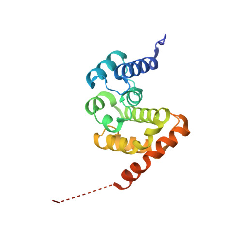
    eIF5, a GTPase-activating protein (GAP) specific for eIF2, plays a critical role in pre-initiation complex assembly and correct AUG selection during eukaryotic translation initiation. eIF5 is involved in the formation of the multifactor complex (MFC), an important intermediate of the 43S pre-initiation complex. The C-terminal domain (CTD) of eIF5 functions as the structural core in the MFC assembly. Here we report the 1.5A crystal structure of eIF5-CTD, confirming that eIF5-CTD contains an atypical HEAT motif. In addition, analyzing the electrostatic potential and the distribution of conserved residues on the protein surface, we confirm and suggest some potential regions of interactions between eIF5-CTD and other eIFs. The structure of eIF5-CTD provides useful information in understanding the mechanism of the MFC assembly.


  • Organizational Affiliation
    • National Laboratory of Biomacromolecules, USTC-IBP Joint Laboratory for Protein Sciences, Institute of Biophysics, Chinese Academy of Sciences, Beijing, People's Republic of China.

Macromolecule Content 

  • Total Structure Weight: 124.4 kDa 
  • Atom Count: 9,151 
  • Modeled Residue Count: 970 
  • Deposited Residue Count: 1,062 
  • Unique protein chains: 1

Macromolecules

Find similar proteins by:|  3D Structure
Entity ID: 1
MoleculeChains  Sequence LengthOrganismDetailsImage
Eukaryotic translation initiation factor 5
A, B, C, D, E
A, B, C, D, E, F
177Saccharomyces cerevisiaeMutation(s): 0 
UniProt
Find proteins for P38431 (Saccharomyces cerevisiae (strain ATCC 204508 / S288c))
Explore P38431 
Go to UniProtKB:  P38431
Entity Groups
Sequence Clusters30% Identity50% Identity70% Identity90% Identity95% Identity100% Identity
UniProt GroupP38431
Sequence Annotations
Expand
Reference Sequence

Small Molecules

Ligands 1 Unique
IDChains Name / Formula / InChI Key2D Diagram3D Interactions
SO4

Query on SO4


Download:Ideal Coordinates CCD File
G [auth A]
H [auth A]
I [auth A]
J [auth B]
K [auth B]
G [auth A],
H [auth A],
I [auth A],
J [auth B],
K [auth B],
L [auth C],
M [auth C],
N [auth D],
O [auth D],
P [auth D],
Q [auth D],
R [auth D],
S [auth E],
T [auth E],
U [auth E],
V [auth E],
W [auth F],
X [auth F],
Y [auth F],
Z [auth F]
SULFATE ION
O4 S
QAOWNCQODCNURD-UHFFFAOYSA-L

Experimental Data & Validation

Experimental Data

  • Method: X-RAY DIFFRACTION
  • Resolution: 1.50 Å
  • R-Value Free:  0.205 (Depositor), 0.204 (DCC) 
  • R-Value Work:  0.177 (Depositor), 0.175 (DCC) 
Space Group: P 1
Unit Cell:
Length ( Å )Angle ( ˚ )
a = 44.849α = 88.6
b = 64.536β = 86.57
c = 108.916γ = 74.93
Software Package:
Software NamePurpose
DENZOdata reduction
SCALEPACKdata scaling
REFMACrefinement
PDB_EXTRACTdata extraction
HKL-2000data reduction
SHARPphasing

Structure Validation

View Full Validation Report


Entry History 

Deposition Data

Revision History  (Full details and data files)

  • Version 1.0: 2006-05-23
    Type: Initial release
  • Version 1.1: 2008-05-01
    Changes: Version format compliance
  • Version 1.2: 2011-07-13
    Changes: Advisory, Version format compliance
  • Version 1.3: 2017-10-18
    Changes: Refinement description
  • Version 1.4: 2024-03-13
    Changes: Data collection, Database references, Derived calculations