2FPD | pdb_00002fpd

Sad structure determination: crystal structure of the intrinsic dimerization sh3 domain of the ib1 scaffold protein


Experimental Data Snapshot

  • Method: X-RAY DIFFRACTION
  • Resolution: 2.05 Å
  • R-Value Free: 
    0.228 (Depositor) 
  • R-Value Work: 
    0.184 (Depositor), 0.194 (DCC) 
  • R-Value Observed: 
    0.184 (Depositor) 

wwPDB Validation 3D Report Full Report

Validation slider image for 2FPD

This is version 2.1 of the entry. See complete history

Literature

A unique set of SH3-SH3 interactions controls IB1 homodimerization

Kristensen, O.Guenat, S.Dar, I.Allaman-Pillet, N.Abderrahmani, A.Ferdaoussi, M.Roduit, R.Maurer, F.Beckmann, J.S.Kastrup, J.S.Gajhede, M.Bonny, C.

(2006) EMBO J 25: 785-797

  • DOI: https://doi.org/10.1038/sj.emboj.7600982
  • Primary Citation Related Structures: 
    2FPD, 2FPE, 2FPF

  • PubMed Abstract: 

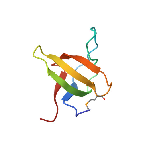
    Islet-brain 1 (IB1 or JIP-1) is a scaffold protein that interacts with components of the c-Jun N-terminal kinase (JNK) signal-transduction pathway. IB1 is expressed at high levels in neurons and in pancreatic beta-cells, where it controls expression of several insulin-secretory components and secretion. IB1 has been shown to homodimerize, but neither the molecular mechanisms nor the function of dimerization have yet been characterized. Here, we show that IB1 homodimerizes through a novel and unique set of Src homology 3 (SH3)-SH3 interactions. X-ray crystallography studies show that the dimer interface covers a region usually engaged in PxxP-mediated ligand recognition, even though the IB1 SH3 domain lacks this motif. The highly stable IB1 homodimer can be significantly destabilized in vitro by three individual point mutations directed against key residues involved in dimerization. Each mutation reduces IB1-dependent basal JNK activity in 293T cells. Impaired dimerization also results in a reduction in glucose transporter type 2 expression and in glucose-dependent insulin secretion in pancreatic beta-cells. Taken together, these results indicate that IB1 homodimerization through its SH3 domain has pleiotropic effects including regulation of the insulin secretion process.


  • Organizational Affiliation
    • Biostructural Research, Department of Medicinal Chemistry, The Danish University of Pharmaceutical Sciences, Copenhagen, Denmark. ok@dfuni.dk

Macromolecule Content 

  • Total Structure Weight: 30.1 kDa 
  • Atom Count: 2,382 
  • Modeled Residue Count: 248 
  • Deposited Residue Count: 248 
  • Unique protein chains: 1

Macromolecules

Find similar proteins by:|  3D Structure
Entity ID: 1
MoleculeChains  Sequence LengthOrganismDetailsImage
C-jun-amino-terminal kinase interacting protein 1
A, B, C, D
62Rattus norvegicusMutation(s): 1 
Gene Names: Mapk8ip1Ib1Jip1
UniProt
Find proteins for Q9R237 (Rattus norvegicus)
Explore Q9R237 
Go to UniProtKB:  Q9R237
Entity Groups
Sequence Clusters30% Identity50% Identity70% Identity90% Identity95% Identity100% Identity
UniProt GroupQ9R237
Sequence Annotations
Expand
Reference Sequence

Oligosaccharides

Help  
Entity ID: 2
MoleculeChains Length2D Diagram GlycosylationD Interactions
alpha-D-glucopyranose-(1-1)-alpha-D-glucopyranose
E
2N/A
Glycosylation Resources
GlyTouCan: G92130SN
GlyCosmos: G92130SN

Small Molecules

Modified Residues  1 Unique
IDChains TypeFormula2D DiagramParent
MSE
Query on MSE
A, B, C, D
L-PEPTIDE LINKINGC5 H11 N O2 SeMET

Biologically Interesting Molecules (External Reference) 

1 Unique

Experimental Data & Validation

Experimental Data

  • Method: X-RAY DIFFRACTION
  • Resolution: 2.05 Å
  • R-Value Free:  0.228 (Depositor) 
  • R-Value Work:  0.184 (Depositor), 0.194 (DCC) 
  • R-Value Observed: 0.184 (Depositor) 
Space Group: P 42 21 2
Unit Cell:
Length ( Å )Angle ( ˚ )
a = 114.486α = 90
b = 114.486β = 90
c = 48.42γ = 90
Software Package:
Software NamePurpose
MOSFLMdata reduction
SCALAdata scaling
SOLVEphasing
SHARPphasing
ARP/wARPmodel building
CCP4model building
CNSrefinement
CCP4data scaling
CCP4phasing

Structure Validation

View Full Validation Report


Entry History 

Deposition Data

Revision History  (Full details and data files)

  • Version 1.0: 2006-02-28
    Type: Initial release
  • Version 1.1: 2008-05-01
    Changes: Version format compliance
  • Version 1.2: 2011-07-13
    Changes: Version format compliance
  • Version 1.3: 2018-05-23
    Changes: Data collection
  • Version 2.0: 2020-07-29
    Type: Remediation
    Reason: Carbohydrate remediation
    Changes: Atomic model, Data collection, Database references, Derived calculations, Non-polymer description, Structure summary
  • Version 2.1: 2024-11-06
    Changes: Data collection, Database references, Structure summary