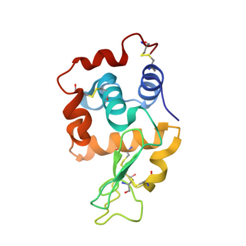
2F2N

Triclinic hen egg lysozyme cross-linked by glutaraldehyde


Experimental Data Snapshot

  • Method: X-RAY DIFFRACTION
  • Resolution: 1.60 Å
  • R-Value Free: 0.169 
  • R-Value Work: 0.137 
  • R-Value Observed: 0.138 

Starting Models: experimental
View more details

wwPDB Validation   3D Report Full Report


This is version 1.4 of the entry. See complete history


Literature

On the edge of the denaturation process: Application of X-ray diffraction to barnase and lysozyme cross-linked crystals with denaturants in molar concentrations.

Salem, M.Mauguen, Y.Prange, T.

(2006) Biochim Biophys Acta 1764: 903-912

  • DOI: https://doi.org/10.1016/j.bbapap.2006.02.009
  • Primary Citation of Related Structures:  
    2F2N, 2F30, 2F4A, 2F4G, 2F4Y, 2F56, 2F5M, 2F5W

  • PubMed Abstract: 

    Structural data about the early step of protein denaturation were obtained from cross-linked crystals for two small proteins: barnase and lysozyme. Several denaturant agents like urea, bromoethanol or thiourea were used at increasing concentrations up to a limit leading to crystal disruption (>or=2 to 6 M). Before the complete destruction of the crystal order started, specific binding sites were observed at the protein surfaces, an indication that the preliminary step of denaturation is the disproportion of intermolecular polar bonds to the benefit of the agent "parasiting" the surface. The analysis of the thermal factors first agree with a stabilization effect at low or moderate concentration of denaturants rapidly followed by a destabilization at specific weak points when the number of sites increase (overflooding effect).


  • Organizational Affiliation

    Université René Descartes, Faculté de pharmacie, Laboratoire de cristallographie et RMN biologiques (UMR-8015, CNRS), 4 av. de l'Observatoire 75270 Paris Cedex 06, France.


Macromolecules
Find similar proteins by:  (by identity cutoff)  |  3D Structure
Entity ID: 1
MoleculeChains Sequence LengthOrganismDetailsImage
Lysozyme C129Gallus gallusMutation(s): 0 
EC: 3.2.1.17
UniProt
Find proteins for P00698 (Gallus gallus)
Explore P00698 
Go to UniProtKB:  P00698
Entity Groups  
Sequence Clusters30% Identity50% Identity70% Identity90% Identity95% Identity100% Identity
UniProt GroupP00698
Sequence Annotations
Expand
  • Reference Sequence
Experimental Data & Validation

Experimental Data

  • Method: X-RAY DIFFRACTION
  • Resolution: 1.60 Å
  • R-Value Free: 0.169 
  • R-Value Work: 0.137 
  • R-Value Observed: 0.138 
  • Space Group: P 1
Unit Cell:
Length ( Å )Angle ( ˚ )
a = 27.17α = 88.67
b = 31.84β = 108.32
c = 34.22γ = 111.87
Software Package:
Software NamePurpose
MOSFLMdata reduction
SCALAdata scaling
SHELXmodel building
SHELXL-97refinement
DENZOdata reduction
SCALEPACKdata scaling

Structure Validation

View Full Validation Report


Entry History 

Deposition Data

Revision History  (Full details and data files)

  • Version 1.0: 2006-04-25
    Type: Initial release
  • Version 1.1: 2008-05-01
    Changes: Version format compliance
  • Version 1.2: 2011-07-13
    Changes: Version format compliance
  • Version 1.3: 2017-10-18
    Changes: Advisory, Refinement description
  • Version 1.4: 2023-08-23
    Changes: Advisory, Data collection, Database references, Derived calculations, Refinement description