2ESL

Human Cyclophilin C in Complex with Cyclosporin A


Experimental Data Snapshot

  • Method: X-RAY DIFFRACTION
  • Resolution: 1.90 Å
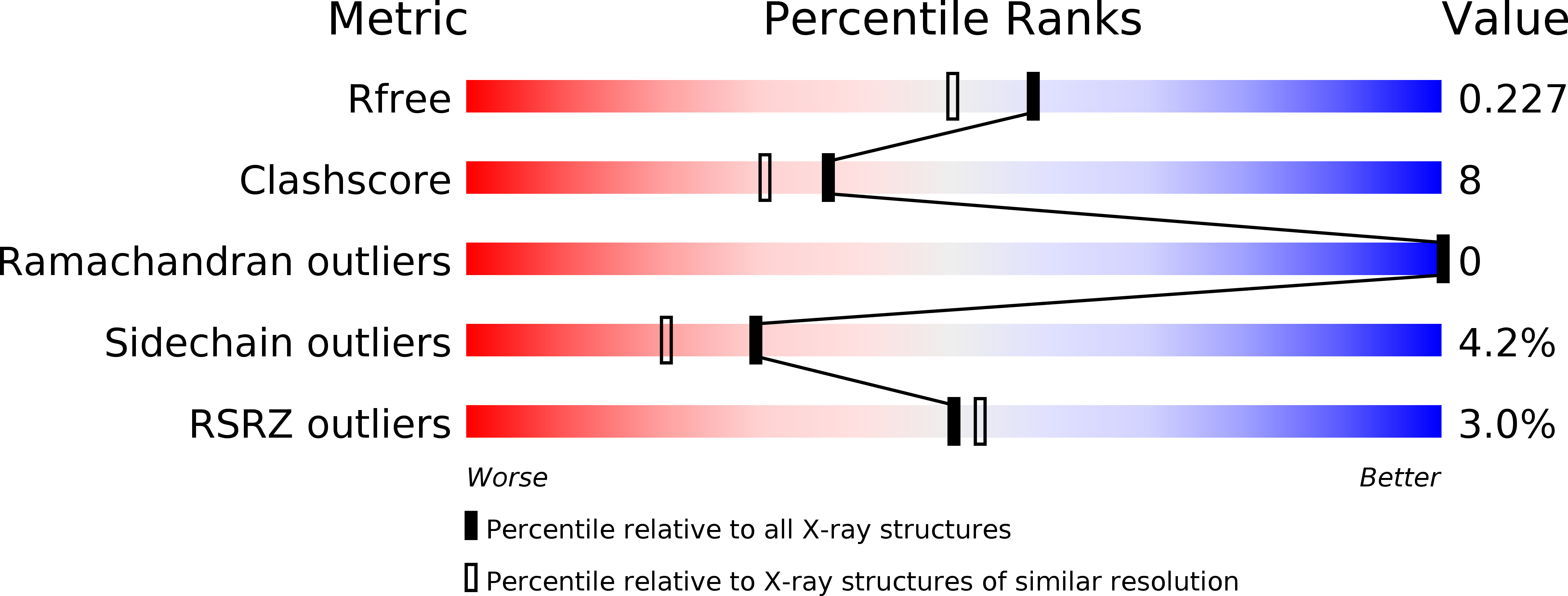
  • R-Value Free: 0.225 
  • R-Value Work: 0.178 
  • R-Value Observed: 0.180 

wwPDB Validation   3D Report Full Report


This is version 1.6 of the entry. See complete history


Literature

Structural and biochemical characterization of the human cyclophilin family of peptidyl-prolyl isomerases.

Davis, T.L.Walker, J.R.Campagna-Slater, V.Finerty, P.J.Paramanathan, R.Bernstein, G.MacKenzie, F.Tempel, W.Ouyang, H.Lee, W.H.Eisenmesser, E.Z.Dhe-Paganon, S.

(2010) PLoS Biol 8: e1000439-e1000439

  • DOI: https://doi.org/10.1371/journal.pbio.1000439
  • Primary Citation of Related Structures:  
    1ZKC, 2ESL, 2GW2, 2HE9, 2HQ6, 2R99

  • PubMed Abstract: 

    Peptidyl-prolyl isomerases catalyze the conversion between cis and trans isomers of proline. The cyclophilin family of peptidyl-prolyl isomerases is well known for being the target of the immunosuppressive drug cyclosporin, used to combat organ transplant rejection. There is great interest in both the substrate specificity of these enzymes and the design of isoform-selective ligands for them. However, the dearth of available data for individual family members inhibits attempts to design drug specificity; additionally, in order to define physiological functions for the cyclophilins, definitive isoform characterization is required. In the current study, enzymatic activity was assayed for 15 of the 17 human cyclophilin isomerase domains, and binding to the cyclosporin scaffold was tested. In order to rationalize the observed isoform diversity, the high-resolution crystallographic structures of seven cyclophilin domains were determined. These models, combined with seven previously solved cyclophilin isoforms, provide the basis for a family-wide structure:function analysis. Detailed structural analysis of the human cyclophilin isomerase explains why cyclophilin activity against short peptides is correlated with an ability to ligate cyclosporin and why certain isoforms are not competent for either activity. In addition, we find that regions of the isomerase domain outside the proline-binding surface impart isoform specificity for both in vivo substrates and drug design. We hypothesize that there is a well-defined molecular surface corresponding to the substrate-binding S2 position that is a site of diversity in the cyclophilin family. Computational simulations of substrate binding in this region support our observations. Our data indicate that unique isoform determinants exist that may be exploited for development of selective ligands and suggest that the currently available small-molecule and peptide-based ligands for this class of enzyme are insufficient for isoform specificity.


  • Organizational Affiliation

    Structural Genomics Consortium, University of Toronto, Toronto, Ontario, Canada.


Macromolecules
Find similar proteins by:  (by identity cutoff)  |  3D Structure
Entity ID: 1
MoleculeChains Sequence LengthOrganismDetailsImage
Peptidyl-prolyl cis-trans isomerase C
A, B, C, D, E
A, B, C, D, E, F
190Homo sapiensMutation(s): 0 
Gene Names: PPICCYPC
EC: 5.2.1.8
UniProt & NIH Common Fund Data Resources
Find proteins for P45877 (Homo sapiens)
Explore P45877 
Go to UniProtKB:  P45877
PHAROS:  P45877
GTEx:  ENSG00000168938 
Entity Groups  
Sequence Clusters30% Identity50% Identity70% Identity90% Identity95% Identity100% Identity
UniProt GroupP45877
Sequence Annotations
Expand
  • Reference Sequence

Find similar proteins by:  Sequence   |   3D Structure  

Entity ID: 2
MoleculeChains Sequence LengthOrganismDetailsImage
CYCLOSPORIN A11Tolypocladium inflatumMutation(s): 0 
Entity Groups  
Sequence Clusters30% Identity50% Identity70% Identity90% Identity95% Identity100% Identity
Sequence Annotations
Expand
  • Reference Sequence
Small Molecules
Ligands 3 Unique
IDChains Name / Formula / InChI Key2D Diagram3D Interactions
SO4
Query on SO4

Download Ideal Coordinates CCD File 
O [auth A]
P [auth B]
Q [auth C]
T [auth D]
U [auth E]
O [auth A],
P [auth B],
Q [auth C],
T [auth D],
U [auth E],
W [auth F]
SULFATE ION
O4 S
QAOWNCQODCNURD-UHFFFAOYSA-L
ZN
Query on ZN

Download Ideal Coordinates CCD File 
N [auth A],
S [auth D]
ZINC ION
Zn
PTFCDOFLOPIGGS-UHFFFAOYSA-N
CA
Query on CA

Download Ideal Coordinates CCD File 
M [auth A],
R [auth D],
V [auth F]
CALCIUM ION
Ca
BHPQYMZQTOCNFJ-UHFFFAOYSA-N
Modified Residues  5 Unique
IDChains TypeFormula2D DiagramParent
ABA
Query on ABA
G [auth I]
H [auth J]
I [auth K]
J [auth L]
K [auth M]
G [auth I],
H [auth J],
I [auth K],
J [auth L],
K [auth M],
L [auth N]
L-PEPTIDE LINKINGC4 H9 N O2ALA
BMT
Query on BMT
G [auth I]
H [auth J]
I [auth K]
J [auth L]
K [auth M]
G [auth I],
H [auth J],
I [auth K],
J [auth L],
K [auth M],
L [auth N]
L-PEPTIDE LINKINGC10 H19 N O3THR
MLE
Query on MLE
G [auth I]
H [auth J]
I [auth K]
J [auth L]
K [auth M]
G [auth I],
H [auth J],
I [auth K],
J [auth L],
K [auth M],
L [auth N]
L-PEPTIDE LINKINGC7 H15 N O2LEU
MVA
Query on MVA
G [auth I]
H [auth J]
I [auth K]
J [auth L]
K [auth M]
G [auth I],
H [auth J],
I [auth K],
J [auth L],
K [auth M],
L [auth N]
L-PEPTIDE LINKINGC6 H13 N O2VAL
SAR
Query on SAR
G [auth I]
H [auth J]
I [auth K]
J [auth L]
K [auth M]
G [auth I],
H [auth J],
I [auth K],
J [auth L],
K [auth M],
L [auth N]
PEPTIDE LINKINGC3 H7 N O2GLY
Biologically Interesting Molecules (External Reference) 1 Unique
Entity ID: 2
IDChains NameType/Class2D Diagram3D Interactions
PRD_000142
Query on PRD_000142
G [auth I]
H [auth J]
I [auth K]
J [auth L]
K [auth M]
G [auth I],
H [auth J],
I [auth K],
J [auth L],
K [auth M],
L [auth N]
Cyclosporin ACyclic peptide / Immunosuppressant
Experimental Data & Validation

Experimental Data

  • Method: X-RAY DIFFRACTION
  • Resolution: 1.90 Å
  • R-Value Free: 0.225 
  • R-Value Work: 0.178 
  • R-Value Observed: 0.180 
  • Space Group: P 21 21 21
Unit Cell:
Length ( Å )Angle ( ˚ )
a = 72.203α = 90
b = 123.802β = 90
c = 135.25γ = 90
Software Package:
Software NamePurpose
PHASERphasing
REFMACrefinement
HKL-2000data reduction
SCALEPACKdata scaling

Structure Validation

View Full Validation Report


Entry History 

Revision History  (Full details and data files)

  • Version 1.0: 2005-12-13
    Type: Initial release
  • Version 1.1: 2011-06-14
    Changes: Version format compliance
  • Version 1.2: 2011-07-13
    Changes: Version format compliance
  • Version 1.3: 2011-07-27
    Changes: Atomic model, Database references, Derived calculations, Non-polymer description, Structure summary
  • Version 1.4: 2012-12-12
    Changes: Other
  • Version 1.5: 2017-06-21
    Changes: Advisory, Database references, Source and taxonomy, Structure summary
  • Version 1.6: 2018-02-07
    Changes: Database references