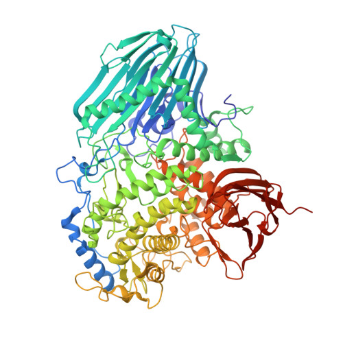
2EAE

Crystal structure of 1,2-a-L-fucosidase from Bifidobacterium bifidum in complexes with products


Experimental Data Snapshot

  • Method: X-RAY DIFFRACTION
  • Resolution: 1.80 Å
  • R-Value Free: 0.219 
  • R-Value Work: 0.180 
  • R-Value Observed: 0.182 

wwPDB Validation   3D Report Full Report


Ligand Structure Quality Assessment 


This is version 2.2 of the entry. See complete history


Literature

Structural basis on the catalytic reaction mechanism of novel 1,2-alpha-L-fucosidase (AFCA) from Bifidobacterium bifidum

Nagae, M.Tsuchiya, A.Katayama, T.Yamamoto, K.Wakatsuki, S.Kato, R.

(2007) J Biol Chem 282: 18497-18509

  • DOI: https://doi.org/10.1074/jbc.M702246200
  • Primary Citation of Related Structures:  
    2EAB, 2EAC, 2EAD, 2EAE

  • PubMed Abstract: 

    1,2-alpha-L-fucosidase (AfcA), which hydrolyzes the glycosidic linkage of Fucalpha1-2Gal via an inverting mechanism, was recently isolated from Bifidobacterium bifidum and classified as the first member of the novel glycoside hydrolase family 95. To better understand the molecular mechanism of this enzyme, we determined the x-ray crystal structures of the AfcA catalytic (Fuc) domain in unliganded and complexed forms with deoxyfuconojirimycin (inhibitor), 2'-fucosyllactose (substrate), and L-fucose and lactose (products) at 1.12-2.10 A resolution. The AfcA Fuc domain is composed of four regions, an N-terminal beta region, a helical linker, an (alpha/alpha)6 helical barrel domain, and a C-terminal beta region, and this arrangement is similar to bacterial phosphorylases. In the complex structures, the ligands were buried in the central cavity of the helical barrel domain. Structural analyses in combination with mutational experiments revealed that the highly conserved Glu566 probably acts as a general acid catalyst. However, no carboxylic acid residue is found at the appropriate position for a general base catalyst. Instead, a water molecule stabilized by Asn423 in the substrate-bound complex is suitably located to perform a nucleophilic attack on the C1 atom of L-fucose moiety in 2'-fucosyllactose, and its location is nearly identical near the O1 atom of beta-L-fucose in the products-bound complex. Based on these data, we propose and discuss a novel catalytic reaction mechanism of AfcA.


  • Organizational Affiliation

    Structural Biology Research Center, Photon Factory, Insititute of Materials Structure Science, High Energy Accelerator Research Organization (KEK), Tsukuba, Ibaraki 305-0801.


Macromolecules
Find similar proteins by:  (by identity cutoff)  |  3D Structure
Entity ID: 1
MoleculeChains Sequence LengthOrganismDetailsImage
Alpha-fucosidase898Bifidobacterium bifidumMutation(s): 1 
EC: 3.2.1.63
UniProt
Find proteins for Q6JV24 (Bifidobacterium bifidum)
Explore Q6JV24 
Go to UniProtKB:  Q6JV24
Entity Groups  
Sequence Clusters30% Identity50% Identity70% Identity90% Identity95% Identity100% Identity
UniProt GroupQ6JV24
Sequence Annotations
Expand
  • Reference Sequence
Oligosaccharides

Help

Entity ID: 2
MoleculeChains Length2D Diagram Glycosylation3D Interactions
beta-D-galactopyranose-(1-4)-alpha-D-glucopyranose
B
2N/A
Glycosylation Resources
GlyTouCan:  G88362QR
GlyCosmos:  G88362QR
GlyGen:  G88362QR
Biologically Interesting Molecules (External Reference) 1 Unique
Entity ID: 2
IDChains NameType/Class2D Diagram3D Interactions
PRD_900008
Query on PRD_900008
B
alpha-lactoseOligosaccharide / Nutrient
Experimental Data & Validation

Experimental Data

  • Method: X-RAY DIFFRACTION
  • Resolution: 1.80 Å
  • R-Value Free: 0.219 
  • R-Value Work: 0.180 
  • R-Value Observed: 0.182 
  • Space Group: P 21 21 21
Unit Cell:
Length ( Å )Angle ( ˚ )
a = 64.519α = 90
b = 70.878β = 90
c = 170.979γ = 90
Software Package:
Software NamePurpose
REFMACrefinement
HKL-2000data collection
HKL-2000data reduction
HKL-2000data scaling
MOLREPphasing

Structure Validation

View Full Validation Report


Ligand Structure Quality Assessment 


Entry History 

Deposition Data

Revision History  (Full details and data files)

  • Version 1.0: 2007-04-24
    Type: Initial release
  • Version 1.1: 2008-04-30
    Changes: Version format compliance
  • Version 1.2: 2011-07-13
    Changes: Version format compliance
  • Version 2.0: 2020-07-29
    Type: Remediation
    Reason: Carbohydrate remediation
    Changes: Atomic model, Data collection, Derived calculations, Non-polymer description, Structure summary
  • Version 2.1: 2021-11-10
    Changes: Database references, Structure summary
  • Version 2.2: 2024-05-29
    Changes: Data collection