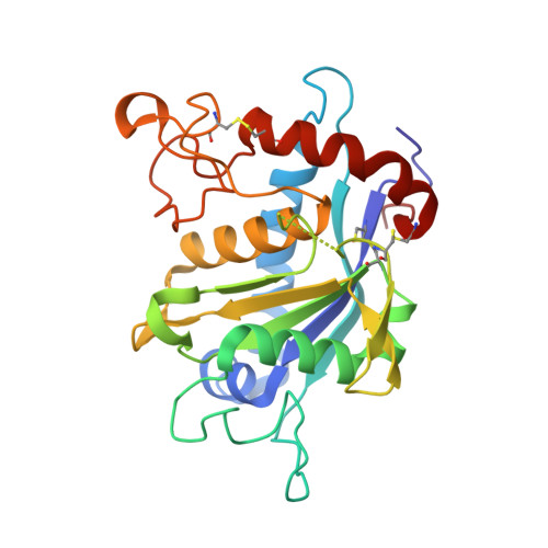
2DDF

Crystal structure of TACE in complex with TAPI-2


Experimental Data Snapshot

  • Method: X-RAY DIFFRACTION
  • Resolution: 1.70 Å
  • R-Value Free: 0.230 
  • R-Value Work: 0.204 
  • R-Value Observed: 0.204 

Starting Model: experimental
View more details

wwPDB Validation   3D Report Full Report


Ligand Structure Quality Assessment 


This is version 1.6 of the entry. See complete history


Literature

Stabilization of the autoproteolysis of TNF-alpha converting enzyme (TACE) results in a novel crystal form suitable for structure-based drug design studies.

Ingram, R.N.Orth, P.Strickland, C.L.Le, H.V.Madison, V.Beyer, B.M.

(2006) Protein Eng Des Sel 19: 155-161

  • DOI: https://doi.org/10.1093/protein/gzj014
  • Primary Citation of Related Structures:  
    2DDF, 2FV9

  • PubMed Abstract: 

    The crystallization of TNF-alpha converting enzyme (TACE) has been useful in understanding the structure-activity relationships of new chemical entities. However, the propensity of TACE to undergo autoproteolysis has made enzyme handling difficult and impeded the identification of inhibitor soakable crystal forms. The autoproteolysis of TACE was found to be specific (Y352-V353) and occurred within a flexible loop that is in close proximity to the P-side of the active site. The rate of autoproteolysis was found to be proportional to the concentration of TACE, suggesting a bimolecular reaction mechanism. A limited specificity study of the S(1)' subsite was conducted using surrogate peptides and suggested substitutions that would stabilize the proteolysis of the loop at positions Y352-V353. Two mutant proteases (V353G and V353S) were generated and proved to be highly resistant to autoproteolysis. The kinetics of the more resistant mutant (V353G) and wild-type TACE were compared and demonstrated virtually identical IC(50) values for a panel of competitive inhibitors. However, the k(cat)/K(m) of the mutant for a larger substrate (P6 - P(6)') was approximately 5-fold lower than that for the wild-type enzyme. Comparison of the complexed wild-type and mutant structures indicated a subtle shift in a peripheral P-side loop (comprising the mutation site) that may be involved in substrate binding/turnover and might explain the mild kinetic difference. The characterization of this stabilized form of TACE has yielded an enzyme with similar native kinetic properties and identified a novel crystal form that is suitable for inhibitor soaking and structure determination.


  • Organizational Affiliation

    Department of Structural Chemistry, Schering-Plough Research Institute, 2015 Galloping Hill Road, Kenilworth, NJ 07033, USA.


Macromolecules
Find similar proteins by:  (by identity cutoff)  |  3D Structure
Entity ID: 1
MoleculeChains Sequence LengthOrganismDetailsImage
ADAM 17
A, B
257Homo sapiensMutation(s): 3 
Gene Names: ADAM17CSVPTACE
EC: 3.4.24.86
UniProt & NIH Common Fund Data Resources
Find proteins for P78536 (Homo sapiens)
Explore P78536 
Go to UniProtKB:  P78536
PHAROS:  P78536
GTEx:  ENSG00000151694 
Entity Groups  
Sequence Clusters30% Identity50% Identity70% Identity90% Identity95% Identity100% Identity
UniProt GroupP78536
Sequence Annotations
Expand
  • Reference Sequence
Small Molecules
Ligands 6 Unique
IDChains Name / Formula / InChI Key2D Diagram3D Interactions
INN
Query on INN

Download Ideal Coordinates CCD File 
E [auth A],
H [auth B]
N-{(2R)-2-[2-(hydroxyamino)-2-oxoethyl]-4-methylpentanoyl}-3-methyl-L-valyl-N-(2-aminoethyl)-L-alaninamide
C19 H37 N5 O5
LMIQCBIEAHJAMZ-GZBFAFLISA-N
CIT
Query on CIT

Download Ideal Coordinates CCD File 
J [auth B]CITRIC ACID
C6 H8 O7
KRKNYBCHXYNGOX-UHFFFAOYSA-N
IMD
Query on IMD

Download Ideal Coordinates CCD File 
I [auth B]IMIDAZOLE
C3 H5 N2
RAXXELZNTBOGNW-UHFFFAOYSA-O
ZN
Query on ZN

Download Ideal Coordinates CCD File 
C [auth A],
G [auth B]
ZINC ION
Zn
PTFCDOFLOPIGGS-UHFFFAOYSA-N
IPA
Query on IPA

Download Ideal Coordinates CCD File 
F [auth A],
K [auth B],
L [auth B],
M [auth B]
ISOPROPYL ALCOHOL
C3 H8 O
KFZMGEQAYNKOFK-UHFFFAOYSA-N
CA
Query on CA

Download Ideal Coordinates CCD File 
D [auth A]CALCIUM ION
Ca
BHPQYMZQTOCNFJ-UHFFFAOYSA-N
Binding Affinity Annotations 
IDSourceBinding Affinity
INN PDBBind:  2DDF IC50: 41 (nM) from 1 assay(s)
Biologically Interesting Molecules (External Reference) 1 Unique
Experimental Data & Validation

Experimental Data

  • Method: X-RAY DIFFRACTION
  • Resolution: 1.70 Å
  • R-Value Free: 0.230 
  • R-Value Work: 0.204 
  • R-Value Observed: 0.204 
  • Space Group: P 21 21 21
Unit Cell:
Length ( Å )Angle ( ˚ )
a = 74.394α = 90
b = 75.639β = 90
c = 103.713γ = 90
Software Package:
Software NamePurpose
REFMACrefinement
PDB_EXTRACTdata extraction

Structure Validation

View Full Validation Report


Ligand Structure Quality Assessment 


Entry History 

Deposition Data

  • Released Date: 2006-03-14 
  • Deposition Author(s): Orth, P.

Revision History  (Full details and data files)

  • Version 1.0: 2006-03-14
    Type: Initial release
  • Version 1.1: 2008-04-30
    Changes: Version format compliance
  • Version 1.2: 2011-07-13
    Changes: Non-polymer description, Version format compliance
  • Version 1.3: 2013-02-27
    Changes: Other
  • Version 1.4: 2021-10-20
    Changes: Database references, Derived calculations
  • Version 1.5: 2023-08-23
    Changes: Data collection, Refinement description
  • Version 1.6: 2024-10-30
    Changes: Structure summary