2CJY | pdb_00002cjy

Extended substrate recognition in caspase-3 revealed by high resolution X-ray structure analysis


Experimental Data Snapshot

  • Method: X-RAY DIFFRACTION
  • Resolution: 1.67 Å
  • R-Value Free: 
    0.206 (Depositor), 0.196 (DCC) 
  • R-Value Work: 
    0.176 (Depositor), 0.167 (DCC) 
  • R-Value Observed: 
    0.176 (Depositor) 

wwPDB Validation 3D Report Full Report

Validation slider image for 2CJY

This is version 1.5 of the entry. See complete history

Literature

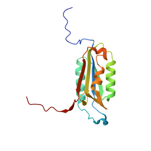
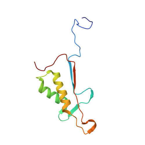
Extended Substrate Recognition in Caspase-3 Revealed by High Resolution X-Ray Structure Analysis

Ganesan, R.Mittl, P.R.E.Jelakovic, S.Grutter, M.G.

(2006) J Mol Biology 359: 1378

  • DOI: https://doi.org/10.1016/j.jmb.2006.04.051
  • Primary Citation Related Structures: 
    2CJX, 2CJY, 2DKO

  • PubMed Abstract: 

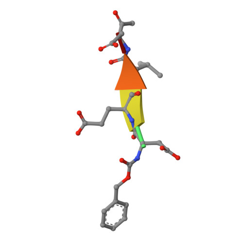
    Caspases are cysteine proteases involved in the signalling cascades of programmed cell death in which caspase-3 plays a central role, since it propagates death signals from intrinsic and extrinsic stimuli to downstream targets. The atomic resolution (1.06 Angstroms) crystal structure of the caspase-3 DEVD-cmk complex reveals the structural basis for substrate selectivity in the S4 pocket. A low-barrier hydrogen bond is observed between the side-chains of the P4 inhibitor aspartic acid and Asp179 of the N-terminal tail of the symmetry related p12 subunit. Site-directed mutagenesis of Asp179 confirmed the significance of this residue in substrate recognition. In the 1.06 Angstroms crystal structure, a radiation damage induced rearrangement of the inhibitor methylketone moiety was observed. The carbon atom that in a substrate would represent the scissile peptide bond carbonyl carbon clearly shows a tetrahedral coordination and resembles the postulated tetrahedral intermediate of the acylation reaction.


  • Organizational Affiliation
    • Biochemisches Institut, Universität Zürich, Switzerland.

Macromolecule Content 

  • Total Structure Weight: 29.28 kDa 
  • Atom Count: 2,376 
  • Modeled Residue Count: 256 
  • Deposited Residue Count: 256 
  • Unique protein chains: 3

Macromolecules

Find similar proteins by:|  3D Structure
Entity ID: 1
MoleculeChains  Sequence LengthOrganismDetailsImage
CASPASE-3147Homo sapiensMutation(s): 0 
Gene Names: CASP3CPP32
EC: 3.4.22.56
UniProt & NIH Common Fund Data Resources
Find proteins for P42574 (Homo sapiens)
Explore P42574 
Go to UniProtKB:  P42574
PHAROS:  P42574
GTEx:  ENSG00000164305 
Entity Groups
Sequence Clusters30% Identity50% Identity70% Identity90% Identity95% Identity100% Identity
UniProt GroupP42574
Sequence Annotations
Expand
Reference Sequence
Find similar proteins by:|  3D Structure
Entity ID: 2
MoleculeChains  Sequence LengthOrganismDetailsImage
CASPASE-3103Homo sapiensMutation(s): 0 
Gene Names: CASP3CPP32
EC: 3.4.22.56
UniProt & NIH Common Fund Data Resources
Find proteins for P42574 (Homo sapiens)
Explore P42574 
Go to UniProtKB:  P42574
PHAROS:  P42574
GTEx:  ENSG00000164305 
Entity Groups
Sequence Clusters30% Identity50% Identity70% Identity90% Identity95% Identity100% Identity
UniProt GroupP42574
Sequence Annotations
Expand
Reference Sequence
Find similar proteins by:  Sequence   |   3D Structure  
Entity ID: 3
MoleculeChains  Sequence LengthOrganismDetailsImage
PHQ-ASP-GLU-VAL-ASP-CHLOROMETHYLKETONEC [auth I]6synthetic constructMutation(s): 0 

Experimental Data & Validation

Experimental Data

  • Method: X-RAY DIFFRACTION
  • Resolution: 1.67 Å
  • R-Value Free:  0.206 (Depositor), 0.196 (DCC) 
  • R-Value Work:  0.176 (Depositor), 0.167 (DCC) 
  • R-Value Observed: 0.176 (Depositor) 
Space Group: I 2 2 2
Unit Cell:
Length ( Å )Angle ( ˚ )
a = 67.468α = 90
b = 83.834β = 90
c = 96.176γ = 90
Software Package:
Software NamePurpose
CNSrefinement
MOSFLMdata reduction
SCALAdata scaling

Structure Validation

View Full Validation Report


Entry History 

Deposition Data

Revision History  (Full details and data files)

  • Version 1.0: 2006-06-27
    Type: Initial release
  • Version 1.1: 2011-07-13
    Changes: Atomic model, Database references, Derived calculations, Non-polymer description, Structure summary, Version format compliance
  • Version 1.2: 2012-11-30
    Changes: Other
  • Version 1.3: 2016-12-21
    Changes: Data collection, Source and taxonomy
  • Version 1.4: 2017-06-28
    Changes: Data collection
  • Version 1.5: 2024-11-20
    Changes: Data collection, Database references, Derived calculations, Other, Structure summary