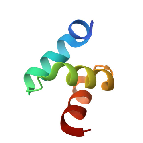
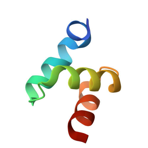
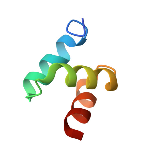
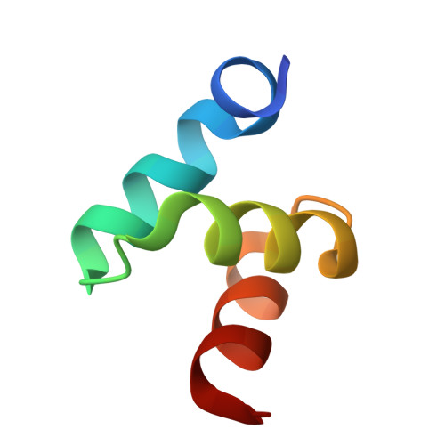
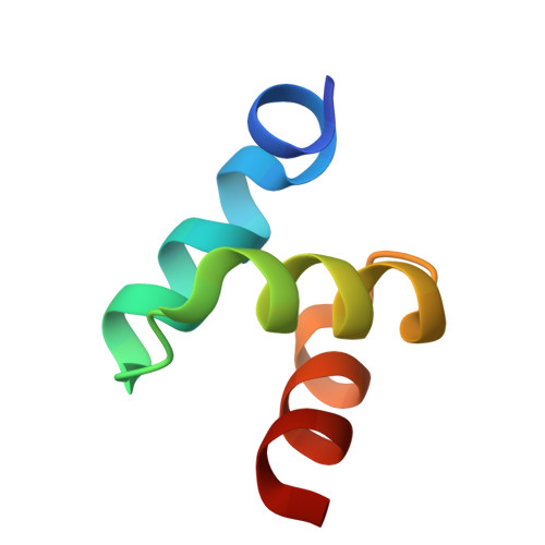
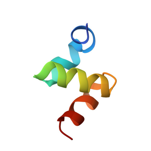
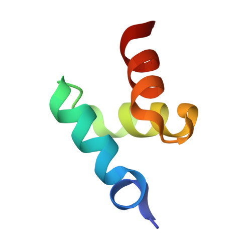
2BWB

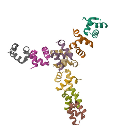
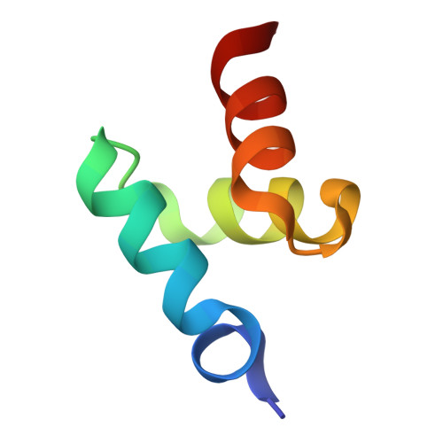
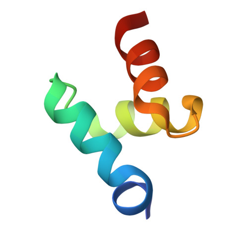
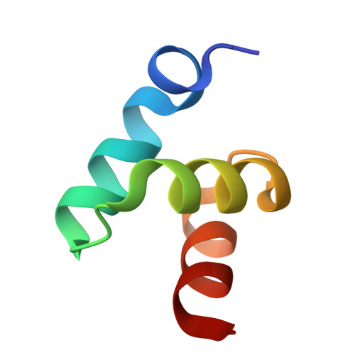
Crystal structure of the UBA domain of Dsk2 from S. cerevisiae


Experimental Data Snapshot

  • Method: X-RAY DIFFRACTION
  • Resolution: 2.30 Å
  • R-Value Free: 0.307 
  • R-Value Work: 0.243 
  • R-Value Observed: 0.246 

wwPDB Validation   3D Report Full Report


This is version 1.3 of the entry. See complete history


Literature

Structures of the Dsk2 Ubl and Uba Domains and Their Complex.

Lowe, E.D.Hasan, N.Trempe, J.-F.Fonso, L.Noble, M.E.M.Endicott, J.A.Johnson, L.N.Brown, N.R.

(2006) Acta Crystallogr D Biol Crystallogr 62: 177

  • DOI: https://doi.org/10.1107/S0907444905037777
  • Primary Citation of Related Structures:  
    2BWB, 2BWE, 2BWF

  • PubMed Abstract: 

    The yeast proteins Dsk2 and Rad23 belong to a family of proteins that contain an N-terminal ubiquitin-like domain (UBL) and a C-terminal ubiquitin-associated domain (UBA). Both Dsk2 and Rad23 function as adaptors to target ubiquitin-labelled proteins to the proteasome through recognition of polyubiquitin (four or more K48-linked ubiquitins) by their UBA domains and to the yeast proteasomal subunit Rpn1 by their UBL domains. The crystal structures of the Dsk2 UBL domain, the Dsk2 UBA domain and the Dsk2 UBA-UBL complex are reported. In the crystal, the Dsk2 UBA domains associate through electrostatic interactions to form ninefold helical ribbons that leave the ubiquitin-binding surface exposed. The UBA-UBL complex explains the reduced affinity of the UBA domain for UBL compared with ubiquitin and has implications for the regulation of Dsk2 adaptor function during ubiquitin-mediated proteasomal targeting. A model is discussed in which two or more Dsk2 UBA molecules may selectively bind to K48-linked polyubiquitin.


  • Organizational Affiliation

    Laboratory of Molecular Biophysics, Department of Biochemistry, University of Oxford, Oxford OX1 3QU, England.


Macromolecules
Find similar proteins by:  (by identity cutoff)  |  3D Structure
Entity ID: 1
MoleculeChains Sequence LengthOrganismDetailsImage
UBIQUITIN-LIKE PROTEIN DSK2
A, B, C, D, E
A, B, C, D, E, F, G, H, I
46Saccharomyces cerevisiaeMutation(s): 0 
UniProt
Find proteins for P48510 (Saccharomyces cerevisiae (strain ATCC 204508 / S288c))
Explore P48510 
Go to UniProtKB:  P48510
Entity Groups  
Sequence Clusters30% Identity50% Identity70% Identity90% Identity95% Identity100% Identity
UniProt GroupP48510
Sequence Annotations
Expand
  • Reference Sequence
Experimental Data & Validation

Experimental Data

  • Method: X-RAY DIFFRACTION
  • Resolution: 2.30 Å
  • R-Value Free: 0.307 
  • R-Value Work: 0.243 
  • R-Value Observed: 0.246 
  • Space Group: C 1 2 1
Unit Cell:
Length ( Å )Angle ( ˚ )
a = 116.311α = 90
b = 44.069β = 114.87
c = 111.525γ = 90
Software Package:
Software NamePurpose
REFMACrefinement
MOSFLMdata reduction
SCALAdata scaling
SHELXphasing

Structure Validation

View Full Validation Report


Entry History 

Deposition Data

Revision History  (Full details and data files)

  • Version 1.0: 2006-01-25
    Type: Initial release
  • Version 1.1: 2011-05-08
    Changes: Version format compliance
  • Version 1.2: 2011-07-13
    Changes: Version format compliance
  • Version 1.3: 2024-05-08
    Changes: Data collection, Database references, Other