2BQZ

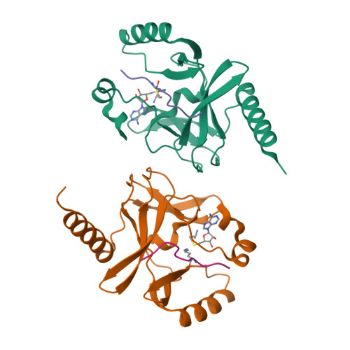
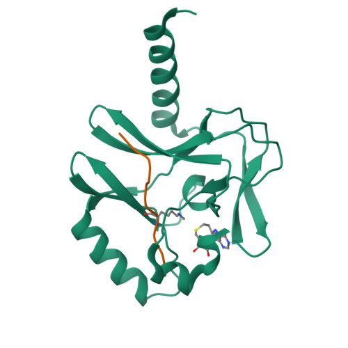
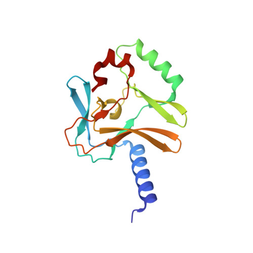
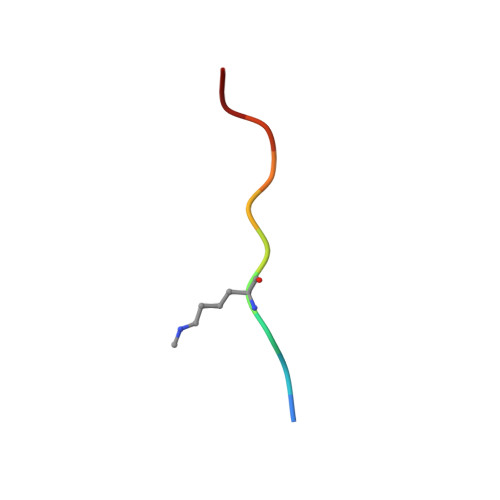
Crystal structure of a ternary complex of the human histone methyltransferase Pr-SET7 (also known as SET8)


Experimental Data Snapshot

  • Method: X-RAY DIFFRACTION
  • Resolution: 1.50 Å
  • R-Value Free: 0.206 
  • R-Value Work: 0.187 
  • R-Value Observed: 0.188 

wwPDB Validation   3D Report Full Report


This is version 1.2 of the entry. See complete history


Literature

Specificity and Mechanism of the Histone Methyltransferase Pr-Set7

Xiao, B.Jing, C.Kelly, G.Walker, P.A.Muskett, F.W.Frenkiel, T.A.Martin, S.R.Sarma, K.Reinberg, D.Gamblin, S.J.Wilson, J.R.

(2005) Genes Dev 19: 1444

  • DOI: https://doi.org/10.1101/gad.1315905
  • Primary Citation of Related Structures:  
    2BQZ

  • PubMed Abstract: 

    Methylation of lysine residues of histones is an important epigenetic mark that correlates with functionally distinct regions of chromatin. We present here the crystal structure of a ternary complex of the enzyme Pr-Set7 (also known as Set8) that methylates Lys 20 of histone H4 (H4-K20). We show that the enzyme is exclusively a mono-methylase and is therefore responsible for a signaling role quite distinct from that established by other enzymes that target this histone residue. We provide evidence from NMR for the C-flanking domains of SET proteins becoming ordered upon addition of AdoMet cofactor and develop a model for the catalytic cycle of these enzymes. The crystal structure reveals the basis of the specificity of the enzyme for H4-K20 because a histidine residue within the substrate, close to the target lysine, is required for completion of the active site. We also show how a highly variable component of the SET domain is responsible for many of the enzymes' interactions with its target histone peptide and probably also how this part of the structure ensures that Pr-Set7 is nucleosome specific.


  • Organizational Affiliation

    National Institute for Medical Research, The Ridgeway, Mill Hill, London, NW7 1AA, United Kingdom.


Macromolecules
Find similar proteins by:  (by identity cutoff)  |  3D Structure
Entity ID: 1
MoleculeChains Sequence LengthOrganismDetailsImage
SET8 PROTEINA,
C [auth E]
161Homo sapiensMutation(s): 0 
EC: 2.1.1.43 (PDB Primary Data), 2.1.1.361 (UniProt), 2.1.1 (UniProt)
UniProt & NIH Common Fund Data Resources
Find proteins for Q9NQR1 (Homo sapiens)
Explore Q9NQR1 
Go to UniProtKB:  Q9NQR1
PHAROS:  Q9NQR1
GTEx:  ENSG00000183955 
Entity Groups  
Sequence Clusters30% Identity50% Identity70% Identity90% Identity95% Identity100% Identity
UniProt GroupQ9NQR1
Sequence Annotations
Expand
  • Reference Sequence

Find similar proteins by:  Sequence   |   3D Structure  

Entity ID: 2
MoleculeChains Sequence LengthOrganismDetailsImage
HISTONE H4B,
D [auth F]
10Homo sapiensMutation(s): 0 
EC: 2.1.1.372
UniProt & NIH Common Fund Data Resources
Find proteins for Q6GMV2 (Homo sapiens)
Explore Q6GMV2 
Go to UniProtKB:  Q6GMV2
PHAROS:  Q6GMV2
GTEx:  ENSG00000135632 
Entity Groups  
Sequence Clusters30% Identity50% Identity70% Identity90% Identity95% Identity100% Identity
UniProt GroupQ6GMV2
Sequence Annotations
Expand
  • Reference Sequence
Small Molecules
Ligands 1 Unique
IDChains Name / Formula / InChI Key2D Diagram3D Interactions
SAH
Query on SAH

Download Ideal Coordinates CCD File 
E [auth A],
F [auth E]
S-ADENOSYL-L-HOMOCYSTEINE
C14 H20 N6 O5 S
ZJUKTBDSGOFHSH-WFMPWKQPSA-N
Modified Residues  1 Unique
IDChains TypeFormula2D DiagramParent
MLZ
Query on MLZ
B,
D [auth F]
L-PEPTIDE LINKINGC7 H16 N2 O2LYS
Experimental Data & Validation

Experimental Data

  • Method: X-RAY DIFFRACTION
  • Resolution: 1.50 Å
  • R-Value Free: 0.206 
  • R-Value Work: 0.187 
  • R-Value Observed: 0.188 
  • Space Group: P 1
Unit Cell:
Length ( Å )Angle ( ˚ )
a = 42.179α = 64.74
b = 46.322β = 86.66
c = 51.998γ = 90.61
Software Package:
Software NamePurpose
REFMACrefinement
DENZOdata reduction
SCALEPACKdata scaling

Structure Validation

View Full Validation Report


Entry History 

Deposition Data

Revision History  (Full details and data files)

  • Version 1.0: 2005-06-08
    Type: Initial release
  • Version 1.1: 2011-05-08
    Changes: Version format compliance
  • Version 1.2: 2011-07-13
    Changes: Version format compliance