2BPD | pdb_00002bpd

STRUCTURE OF MURINE DECTIN-1


Experimental Data Snapshot

  • Method: X-RAY DIFFRACTION
  • Resolution: 1.50 Å
  • R-Value Free: 
    0.270 (Depositor), 0.179 (DCC) 
  • R-Value Work: 
    0.214 (Depositor), 0.215 (DCC) 
  • R-Value Observed: 
    0.216 (Depositor) 

Starting Models: experimental
View more details

wwPDB Validation 3D Report Full Report

Validation slider image for 2BPD

This is version 1.6 of the entry. See complete history

Literature

Structure of the Fungal Beta-Glucan-Binding Immune Receptor Dectin-1: Implications for Function.

Brown, J.O'Callaghan, C.A.Marshall, A.S.J.Gilbert, R.J.C.Siebold, C.Gordon, S.Brown, G.D.Jones, E.Y.

(2007) Protein Sci 16: 1042

  • DOI: https://doi.org/10.1110/ps.072791207
  • Primary Citation Related Structures: 
    2BPD, 2BPE, 2BPH, 2CL8

  • PubMed Abstract: 

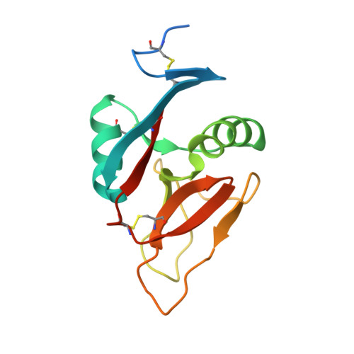
    The murine molecule dectin-1 (known as the beta-glucan receptor in humans) is an immune cell surface receptor implicated in the immunological defense against fungal pathogens. Sequence analysis has indicated that the dectin-1 extracellular domain is a C-type lectin-like domain, and functional studies have established that it binds fungal beta-glucans. We report several dectin-1 crystal structures, including a high-resolution structure and a 2.8 angstroms resolution structure in which a short soaked natural beta-glucan is trapped in the crystal lattice. In vitro characterization of dectin-1 in the presence of its natural ligand indicates higher-order complex formation between dectin-1 and beta-glucans. These combined structural and biophysical data considerably extend the current knowledge of dectin-1 structure and function, and suggest potential mechanisms of defense against fungal pathogens.


  • Organizational Affiliation
    • CR-UK Receptor Structure Research Group, Division of Structural Biology, Wellcome Trust Centre for Human Genetics, University of Oxford, Oxford, OX3 7BN, United Kingdom.

Macromolecule Content 

  • Total Structure Weight: 32.66 kDa 
  • Atom Count: 2,545 
  • Modeled Residue Count: 256 
  • Deposited Residue Count: 284 
  • Unique protein chains: 1

Macromolecules

Find similar proteins by:|  3D Structure
Entity ID: 1
MoleculeChains  Sequence LengthOrganismDetailsImage
DECTIN-1
A, B
142Mus musculusMutation(s): 0 
UniProt
Find proteins for Q6QLQ4 (Mus musculus)
Explore Q6QLQ4 
Go to UniProtKB:  Q6QLQ4
Entity Groups
Sequence Clusters30% Identity50% Identity70% Identity90% Identity95% Identity100% Identity
UniProt GroupQ6QLQ4
Sequence Annotations
Expand
Reference Sequence

Experimental Data & Validation

Experimental Data

  • Method: X-RAY DIFFRACTION
  • Resolution: 1.50 Å
  • R-Value Free:  0.270 (Depositor), 0.179 (DCC) 
  • R-Value Work:  0.214 (Depositor), 0.215 (DCC) 
  • R-Value Observed: 0.216 (Depositor) 
Space Group: P 32
Unit Cell:
Length ( Å )Angle ( ˚ )
a = 57.104α = 90
b = 57.104β = 90
c = 73.422γ = 120
Software Package:
Software NamePurpose
DENZOdata reduction
SCALEPACKdata scaling
CaspRphasing
AMoREphasing
REFMACrefinement

Structure Validation

View Full Validation Report


Entry History 

Deposition Data

Revision History  (Full details and data files)

  • Version 1.0: 2006-08-30
    Type: Initial release
  • Version 1.1: 2011-07-13
    Changes: Advisory, Version format compliance
  • Version 1.2: 2017-07-12
    Changes: Refinement description
  • Version 1.3: 2019-04-03
    Changes: Data collection, Source and taxonomy
  • Version 1.4: 2020-03-25
    Changes: Derived calculations, Other
  • Version 1.5: 2023-12-13
    Changes: Data collection, Database references, Refinement description
  • Version 1.6: 2024-11-13
    Changes: Structure summary