2BNN | pdb_00002bnn

The structure of Hydroxypropylphosphonic acid epoxidase from S. wedmorenis in complex with fosfomycin


Experimental Data Snapshot

  • Method: X-RAY DIFFRACTION
  • Resolution: 2.50 Å
  • R-Value Free: 
    0.270 (Depositor) 
  • R-Value Work: 
    0.184 (Depositor), 0.186 (DCC) 
  • R-Value Observed: 
    0.188 (Depositor) 

wwPDB Validation 3D Report Full Report

Validation slider image for 2BNN

This is version 1.4 of the entry. See complete history

Literature

Structure and Reactivity of Hydroxypropylphosphonic Acid Epoxidase in Fosfomycin Biosynthesis by a Cation- and Flavin-Dependent Mechanism.

Mcluskey, K.Cameron, S.Hammerschmidt, F.Hunter, W.N.

(2005) Proc Natl Acad Sci U S A 102: 14221

  • DOI: https://doi.org/10.1073/pnas.0504314102
  • Primary Citation Related Structures: 
    2BNM, 2BNN, 2BNO

  • PubMed Abstract: 

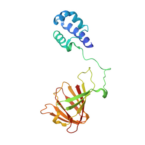
    The biosynthesis of fosfomycin, an oxirane antibiotic in clinical use, involves a unique epoxidation catalyzed by (S)-2-hydroxypropylphosphonic acid epoxidase (HPPE). The reaction is essentially dehydrogenation of a secondary alcohol. A high-resolution crystallographic analysis reveals that the HPPE subunit displays a two-domain combination. The C-terminal or catalytic domain has the cupin fold that binds a divalent cation, whereas the N-terminal domain carries a helix-turn-helix motif with putative DNA-binding helices positioned 34 A apart. The structure of HPPE serves as a model for numerous proteins, of ill-defined function, predicted to be transcription factors but carrying a cupin domain at the C terminus. Structure-reactivity analyses reveal conformational changes near the catalytic center driven by the presence or absence of ligand, that HPPE is a Zn(2+)/Fe(2+)-dependent epoxidase, proof that flavin mononucleotide is required for catalysis, and allow us to propose a simple mechanism that is compatible with previous experimental data. The participation of the redox inert Zn(2+) in the mechanism is surprising and indicates that Lewis acid properties of the metal ions are sufficient to polarize the substrate and, aided by flavin mononucleotide reduction, facilitate the epoxidation.


  • Organizational Affiliation
    • Division of Biological Chemistry and Molecular Microbiology, School of Life Sciences, University of Dundee, Dundee DD1 5EH, United Kingdom.

Macromolecule Content 

  • Total Structure Weight: 43.13 kDa 
  • Atom Count: 3,223 
  • Modeled Residue Count: 383 
  • Deposited Residue Count: 396 
  • Unique protein chains: 1

Macromolecules

Find similar proteins by:|  3D Structure
Entity ID: 1
MoleculeChains  Sequence LengthOrganismDetailsImage
EPOXIDASE
A, B
198Streptomyces wedmorensisMutation(s): 0 
EC: 1.11.1.23
UniProt
Find proteins for Q56185 (Streptomyces wedmorensis)
Explore Q56185 
Go to UniProtKB:  Q56185
Entity Groups
Sequence Clusters30% Identity50% Identity70% Identity90% Identity95% Identity100% Identity
UniProt GroupQ56185
Sequence Annotations
Expand
Reference Sequence

Experimental Data & Validation

Experimental Data

  • Method: X-RAY DIFFRACTION
  • Resolution: 2.50 Å
  • R-Value Free:  0.270 (Depositor) 
  • R-Value Work:  0.184 (Depositor), 0.186 (DCC) 
  • R-Value Observed: 0.188 (Depositor) 
Space Group: P 65 2 2
Unit Cell:
Length ( Å )Angle ( ˚ )
a = 85.602α = 90
b = 85.602β = 90
c = 217.989γ = 120
Software Package:
Software NamePurpose
REFMACrefinement
DENZOdata reduction
SCALEPACKdata scaling

Structure Validation

View Full Validation Report


Entry History 

Deposition Data

Revision History  (Full details and data files)

  • Version 1.0: 2005-10-05
    Type: Initial release
  • Version 1.1: 2011-05-08
    Changes: Version format compliance
  • Version 1.2: 2011-07-13
    Changes: Version format compliance
  • Version 1.3: 2017-06-28
    Changes: Data collection
  • Version 1.4: 2024-05-08
    Changes: Data collection, Database references, Derived calculations, Other