23FX | pdb_000023fx

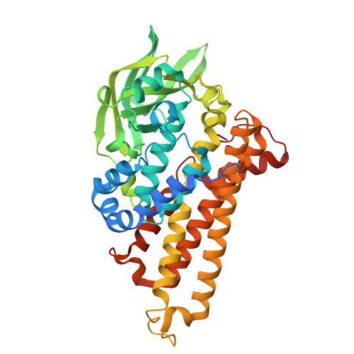
Crystal structure of sulX mutant-A399S in complex with FMN and sulfamethazine


Experimental Data Snapshot

  • Method: X-RAY DIFFRACTION
  • Resolution: 2.47 Å
  • R-Value Free: 
    0.246 (Depositor), 0.250 (DCC) 
  • R-Value Work: 
    0.184 (Depositor), 0.197 (DCC) 
  • R-Value Observed: 
    0.187 (Depositor) 

Starting Model: in silico
View more details

wwPDB Validation 3D Report Full Report

Validation slider image for 23FX

Ligand Structure Quality Assessment 


This is version 1.0 of the entry. See complete history. 

Literature

Structural insights into sulfonamide degradation by a two-component flavin-dependent monooxygenase.

Hu, Y., Liu, W., Zhang, Q., Gao, Z., Zhang, H., Li, H., Dai, L.

(2026) J Hazard Mater 517: 143643-143643

  • DOI: https://doi.org/10.1016/j.jhazmat.2026.143643
  • Primary Citation Related Structures: 
    21CF, 21CG, 21CV, 21DW, 21GB, 21OB, 23FX, 23FY, 9XT7, 9XVK

  • PubMed Abstract: 

    The sulfonamide-degrading monooxygenase sulX plays a dual role in bioremediation and antibiotic resistance, yet its molecular mechanism remains elusive. Here we report crystal structures of sulX in its ligand-free form, as an FMN-bound binary complex, and as ternary complexes with six distinct sulfonamides at resolutions ranging from 2.09 to 3.03 Å. These structures reveal that sulfonamides bind on the re face of the flavin isoalloxazine ring, with their conserved 4-aminophenol and sulfonyl moieties tightly anchored by π-stacking and hydrogen-bonding interactions, while the divergent aminated substituents extend into an open cleft with minimal specific contacts-a feature that explains the enzyme's remarkable capacity to accommodate diverse sulfonamide substrates. By integrating structural and biochemical data, we propose a catalytic mechanism involving F265-mediated substrate gating and H397-facilitated proton transfer. In addition, structure-based engineering yielded enhanced variants: A399S, which introduces new hydrogen bonds, showed a 37% increase in activity, and P422K achieved a 29% improvement. Our findings establish the structural basis of sulX-mediated sulfonamide degradation and provide a foundation for understanding sulfonamide resistance mechanisms.


  • Organizational Affiliation: 
    • State Key Laboratory of Biocatalysis and Enzyme Engineering, Hubei Hongshan Laboratory, Hubei Key Laboratory of Industrial Biotechnology, School of Life Sciences, Hubei University, Wuhan 430062, PR China.

Macromolecule Content 

  • Total Structure Weight: 47.21 kDa 
  • Atom Count: 3,270 
  • Modeled Residue Count: 394 
  • Deposited Residue Count: 423 
  • Unique protein chains: 1

Macromolecules

Find similar proteins by:|  3D Structure
Entity ID: 1
MoleculeChains  Sequence LengthOrganismDetailsImage
Sulfonamide monooxygenase423Microbacterium sp. CJ77Mutation(s): 1 
Gene Names: sulX
UniProt
Find proteins for A0A482P9Z9 (Microbacterium sp. CJ77)
Explore A0A482P9Z9 
Go to UniProtKB:  A0A482P9Z9
Entity Groups
Sequence Clusters30% Identity50% Identity70% Identity90% Identity95% Identity100% Identity
UniProt GroupA0A482P9Z9
Sequence Annotations
Expand
Reference Sequence

Small Molecules

Ligands 5 Unique
IDChains Name / Formula / InChI Key2D Diagram3D Interactions
FMN
(Subject of Investigation/LOI)

Query on FMN


Download:Ideal Coordinates CCD File
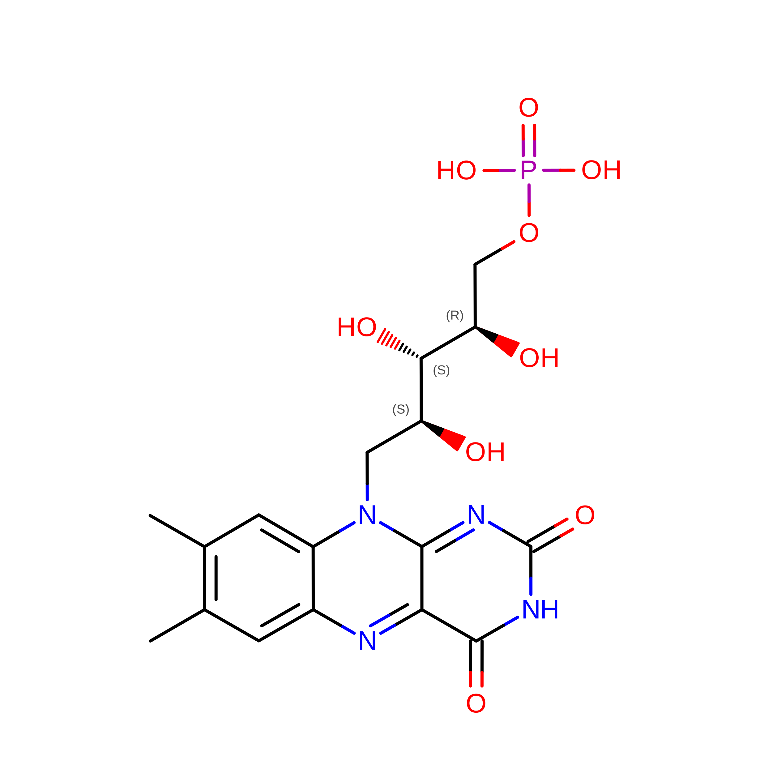
B [auth A]FLAVIN MONONUCLEOTIDE
C17 H21 N4 O9 P
FVTCRASFADXXNN-SCRDCRAPSA-N
A1E1F(
Subject of Investigation/LOI)

Query on A1E1F


Download:Ideal Coordinates CCD File
C [auth A]Sulfamethazine
C12 H14 N4 O2 S
ASWVTGNCAZCNNR-UHFFFAOYSA-N
PEG

Query on PEG


Download:Ideal Coordinates CCD File
F [auth A]DI(HYDROXYETHYL)ETHER
C4 H10 O3
MTHSVFCYNBDYFN-UHFFFAOYSA-N
SO4

Query on SO4


Download:Ideal Coordinates CCD File
D [auth A],
E [auth A]
SULFATE ION
O4 S
QAOWNCQODCNURD-UHFFFAOYSA-L
ZN

Query on ZN


Download:Ideal Coordinates CCD File
G [auth A],
H [auth A],
I [auth A]
ZINC ION
Zn
PTFCDOFLOPIGGS-UHFFFAOYSA-N

Experimental Data & Validation

Experimental Data

  • Method: X-RAY DIFFRACTION
  • Resolution: 2.47 Å
  • R-Value Free:  0.246 (Depositor), 0.250 (DCC) 
  • R-Value Work:  0.184 (Depositor), 0.197 (DCC) 
  • R-Value Observed: 0.187 (Depositor) 
Space Group: P 62 2 2
Unit Cell:
Length ( Å )Angle ( ˚ )
a = 133.72α = 90
b = 133.72β = 90
c = 138.92γ = 120
Software Package:
Software NamePurpose
REFMACrefinement
PDB_EXTRACTdata extraction
SAINTdata reduction
SAINTdata scaling
PHASERphasing

Structure Validation

View Full Validation Report


Ligand Structure Quality Assessment 


Entry History 

& Funding Information

Deposition Data


Funding OrganizationLocationGrant Number
Ministry of Science and Technology (MoST, China)China2021YFC2100400

Revision History  (Full details and data files)

  • Version 1.0: 2026-09-30
    Type: Initial release