1V7W | pdb_00001v7w

Crystal structure of Vibrio proteolyticus chitobiose phosphorylase in complex with GlcNAc


Experimental Data Snapshot

  • Method: X-RAY DIFFRACTION
  • Resolution: 1.60 Å
  • R-Value Free: 
    0.179 (Depositor), 0.149 (DCC) 
  • R-Value Work: 
    0.157 (Depositor), 0.155 (DCC) 
  • R-Value Observed: 
    0.157 (Depositor) 

wwPDB Validation 3D Report Full Report

Validation slider image for 1V7W

Ligand Structure Quality Assessment 


This is version 2.1 of the entry. See complete history

Literature

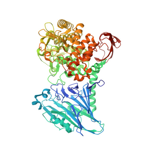
Chitobiose phosphorylase from Vibrio proteolyticus, a member of glycosyl transferase family 36, has a clan GH-L-like (alpha/alpha)(6) barrel fold.

Hidaka, M.Honda, Y.Kitaoka, M.Nirasawa, S.Hayashi, K.Wakagi, T.Shoun, H.Fushinobu, S.

(2004) Structure 12: 937-947

  • DOI: https://doi.org/10.1016/j.str.2004.03.027
  • Primary Citation Related Structures: 
    1V7V, 1V7W, 1V7X

  • PubMed Abstract: 

    Vibrio proteolyticus chitobiose phosphorylase (ChBP) belongs to glycosyl transferase family 36 (GT-36), and catalyzes the reversible phosphorolysis of chitobiose into alpha-GlcNAc-1-phosphate and GlcNAc with inversion of the anomeric configuration. As the first known structures of a GT-36 enzyme, we determined the crystal structure of ChBP in a ternary complex with GlcNAc and SO(4). It is also the first structures of an inverting phosphorolytic enzyme in a complex with a sugar and a sulfate ion, and reveals a pseudo-ternary complex structure of enzyme-sugar-phosphate. ChBP comprises a beta sandwich domain and an (alpha/alpha)(6) barrel domain, constituting a distinctive structure among GT families. Instead, it shows significant structural similarity with glycoside hydrolase (GH) enzymes, glucoamylases (GH-15), and maltose phosphorylase (GH-65) in clan GH-L. The structural similarity reported here, together with distant sequence similarities between ChBP and GHs, led to the reclassification of family GT-36 into a novel GH family, namely GH-94.


  • Organizational Affiliation
    • Department of Biotechnology, The University of Tokyo, 1-1-1, Yayoi, Bunkyo-ku, Tokyo 113-8657, Japan.

Macromolecule Content 

  • Total Structure Weight: 91.88 kDa 
  • Atom Count: 7,168 
  • Modeled Residue Count: 779 
  • Deposited Residue Count: 807 
  • Unique protein chains: 1

Macromolecules

Find similar proteins by:|  3D Structure
Entity ID: 1
MoleculeChains  Sequence LengthOrganismDetailsImage
chitobiose phosphorylase807Vibrio proteolyticusMutation(s): 0 
Gene Names: chbp
EC: 2.4.1 (PDB Primary Data), 2.4.1.280 (UniProt)
UniProt
Find proteins for Q76IQ9 (Vibrio proteolyticus)
Explore Q76IQ9 
Go to UniProtKB:  Q76IQ9
Entity Groups
Sequence Clusters30% Identity50% Identity70% Identity90% Identity95% Identity100% Identity
UniProt GroupQ76IQ9
Sequence Annotations
Expand
Reference Sequence

Small Molecules

Ligands 4 Unique
IDChains Name / Formula / InChI Key2D Diagram3D Interactions
NAG

Query on NAG


Download:Ideal Coordinates CCD File
C [auth A]2-acetamido-2-deoxy-beta-D-glucopyranose
C8 H15 N O6
OVRNDRQMDRJTHS-FMDGEEDCSA-N
NDG

Query on NDG


Download:Ideal Coordinates CCD File
B [auth A]2-acetamido-2-deoxy-alpha-D-glucopyranose
C8 H15 N O6
OVRNDRQMDRJTHS-PVFLNQBWSA-N
CA

Query on CA


Download:Ideal Coordinates CCD File
D [auth A],
E [auth A],
F [auth A]
CALCIUM ION
Ca
BHPQYMZQTOCNFJ-UHFFFAOYSA-N
CL

Query on CL


Download:Ideal Coordinates CCD File
G [auth A]CHLORIDE ION
Cl
VEXZGXHMUGYJMC-UHFFFAOYSA-M

Experimental Data & Validation

Experimental Data

  • Method: X-RAY DIFFRACTION
  • Resolution: 1.60 Å
  • R-Value Free:  0.179 (Depositor), 0.149 (DCC) 
  • R-Value Work:  0.157 (Depositor), 0.155 (DCC) 
  • R-Value Observed: 0.157 (Depositor) 
Space Group: C 1 2 1
Unit Cell:
Length ( Å )Angle ( ˚ )
a = 141.064α = 90
b = 70.729β = 98.41
c = 80.41γ = 90
Software Package:
Software NamePurpose
CNSrefinement
HKL-2000data reduction
SCALEPACKdata scaling
SOLVEphasing

Structure Validation

View Full Validation Report


Ligand Structure Quality Assessment 


Entry History 

Deposition Data

Revision History  (Full details and data files)

  • Version 1.0: 2004-06-22
    Type: Initial release
  • Version 1.1: 2008-04-27
    Changes: Version format compliance
  • Version 1.2: 2011-07-13
    Changes: Derived calculations, Version format compliance
  • Version 2.0: 2020-07-29
    Type: Remediation
    Reason: Carbohydrate remediation
    Changes: Atomic model, Data collection, Database references, Derived calculations, Structure summary
  • Version 2.1: 2023-12-27
    Changes: Data collection, Database references, Structure summary