1Z3F | pdb_00001z3f

Structure of ellipticine in complex with a 6-bp DNA


Experimental Data Snapshot

  • Method: X-RAY DIFFRACTION
  • Resolution: 1.50 Å
  • R-Value Free: 
    0.242 (Depositor), 0.256 (DCC) 
  • R-Value Work: 
    0.220 (Depositor), 0.223 (DCC) 
  • R-Value Observed: 
    0.222 (Depositor) 

wwPDB Validation 3D Report Full Report

Validation slider image for 1Z3F

Ligand Structure Quality Assessment 


This is version 1.4 of the entry. See complete history

Literature

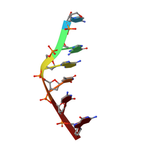
The anticancer agent ellipticine unwinds DNA by intercalative binding in an orientation parallel to base pairs.

Canals, A.Purciolas, M.Aymami, J.Coll, M.

(2005) Acta Crystallogr D Biol Crystallogr 61: 1009-1012

  • DOI: https://doi.org/10.1107/S0907444905015404
  • Primary Citation Related Structures: 
    1Z3F

  • PubMed Abstract: 

    Ellipticine is a natural plant product that has been found to be a powerful anticancer drug. Although still unclear, its mechanism of action is considered to be mainly based on DNA intercalation and/or the inhibition of topoisomerase II. Many experimental data suggest an intercalation based on stacking interactions along the major base-pair axis, but alternative binding modes have been proposed, in particular for ellipticine derivatives. The 1.5 A resolution structure of ellipticine complexed to a 6 bp oligonucleotide unveils its mode of binding and enables a detailed analysis of the distorting effects of the drug on the DNA.


  • Organizational Affiliation
    • Institut de Biologia Molecular de Barcelona, Consejo Superior de Investigaciones Científicas, Parc Científic de Barcelona, Baldiri Reixac 10-12, 08028 Barcelona, Spain. acpcri@ibmb.csic.es

Macromolecule Content 

  • Total Structure Weight: 4.29 kDa 
  • Atom Count: 308 
  • Modeled Residue Count: 12 
  • Deposited Residue Count: 12 
  • Unique nucleic acid chains: 1

Macromolecules

Find similar nucleic acids by:  Sequence
Entity ID: 1
MoleculeChains LengthOrganismImage
5'-D(*CP*GP*AP*TP*CP*G)-3'
A, B
6N/A
Sequence Annotations
Expand
Reference Sequence

Experimental Data & Validation

Experimental Data

  • Method: X-RAY DIFFRACTION
  • Resolution: 1.50 Å
  • R-Value Free:  0.242 (Depositor), 0.256 (DCC) 
  • R-Value Work:  0.220 (Depositor), 0.223 (DCC) 
  • R-Value Observed: 0.222 (Depositor) 
Space Group: P 65
Unit Cell:
Length ( Å )Angle ( ˚ )
a = 24.86α = 90
b = 24.86β = 90
c = 78.86γ = 120
Software Package:
Software NamePurpose
REFMACrefinement
ProDCdata collection
XDSdata scaling
AMoREphasing

Structure Validation

View Full Validation Report


Ligand Structure Quality Assessment 


Entry History 

Deposition Data

Revision History  (Full details and data files)

  • Version 1.0: 2005-07-19
    Type: Initial release
  • Version 1.1: 2008-04-30
    Changes: Version format compliance
  • Version 1.2: 2011-07-13
    Changes: Version format compliance
  • Version 1.3: 2017-10-11
    Changes: Refinement description
  • Version 1.4: 2024-02-14
    Changes: Data collection, Database references, Derived calculations