1YS2 | pdb_00001ys2

Burkholderia cepacia lipase complexed with hexylphosphonic acid (S) 2-methyl-3-phenylpropyl ester


Experimental Data Snapshot

  • Method: X-RAY DIFFRACTION
  • Resolution: 1.50 Å
  • R-Value Free: 
    0.163 (Depositor), 0.168 (DCC) 
  • R-Value Work: 
    0.151 (Depositor), 0.157 (DCC) 
  • R-Value Observed: 
    0.151 (Depositor) 

Starting Model: experimental
View more details

wwPDB Validation 3D Report Full Report

Validation slider image for 1YS2

Ligand Structure Quality Assessment 


This is version 1.4 of the entry. See complete history

Literature

Mirror-Image Packing in Enantiomer Discrimination Molecular Basis for the Enantioselectivity of B.cepacia Lipase toward 2-Methyl-3-Phenyl-1-Propanol.

Mezzetti, A.Schrag, J.D.Cheong, C.S.Kazlauskas, R.J.

(2005) Chem Biol 12: 427-437

  • DOI: https://doi.org/10.1016/j.chembiol.2005.01.016
  • Primary Citation Related Structures: 
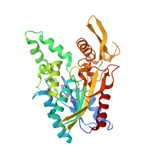
    1YS1, 1YS2

  • PubMed Abstract: 

    Synthetic chemists often exploit the high enantioselectivity of lipases to prepare pure enantiomers of primary alcohols, but the molecular basis for this enantioselectivity is unknown. The crystal structures of two phosphonate transition-state analogs bound to Burkholderia cepacia lipase reveal this molecular basis for a typical primary alcohol: 2-methyl-3-phenyl-1-propanol. The enantiomeric alcohol moieties adopt surprisingly similar orientations, with only subtle differences that make it difficult to predict how to alter enantioselectivity. These structures, along with a survey of previous structures of enzyme bound enantiomers, reveal that binding of enantiomers does not involve an exchange of two substituent positions as most researchers assumed. Instead, the enantiomers adopt mirror-image packing, where three of the four substituents at the stereocenter lie in similar positions. The fourth substituent, hydrogen, points in opposite directions.


  • Organizational Affiliation
    • Department of Chemistry, McGill University, Montréal, Québec H3A 2K6, Canada.

Macromolecule Content 

  • Total Structure Weight: 33.4 kDa 
  • Atom Count: 2,498 
  • Modeled Residue Count: 320 
  • Deposited Residue Count: 320 
  • Unique protein chains: 1

Macromolecules

Find similar proteins by:|  3D Structure
Entity ID: 1
MoleculeChains  Sequence LengthOrganismDetailsImage
LipaseA [auth X]320Burkholderia cepaciaMutation(s): 0 
EC: 3.1.1.3
UniProt
Find proteins for P22088 (Burkholderia cepacia)
Explore P22088 
Go to UniProtKB:  P22088
Entity Groups
Sequence Clusters30% Identity50% Identity70% Identity90% Identity95% Identity100% Identity
UniProt GroupP22088
Sequence Annotations
Expand
Reference Sequence

Small Molecules

Ligands 2 Unique
IDChains Name / Formula / InChI Key2D Diagram3D Interactions
2HS

Query on 2HS


Download:Ideal Coordinates CCD File
C [auth X]HEXYLPHOSPHONIC ACID (S)-2-METHYL-3-PHENYLPROPYL ESTER
C16 H27 O3 P
MMTDYBZZRYOMFD-HNNXBMFYSA-N
CA

Query on CA


Download:Ideal Coordinates CCD File
B [auth X]CALCIUM ION
Ca
BHPQYMZQTOCNFJ-UHFFFAOYSA-N

Experimental Data & Validation

Experimental Data

  • Method: X-RAY DIFFRACTION
  • Resolution: 1.50 Å
  • R-Value Free:  0.163 (Depositor), 0.168 (DCC) 
  • R-Value Work:  0.151 (Depositor), 0.157 (DCC) 
  • R-Value Observed: 0.151 (Depositor) 
Space Group: C 1 2 1
Unit Cell:
Length ( Å )Angle ( ˚ )
a = 88.93α = 90
b = 46.233β = 121.35
c = 84.729γ = 90
Software Package:
Software NamePurpose
REFMACrefinement
DENZOdata reduction
SCALEPACKdata scaling
CNSphasing

Structure Validation

View Full Validation Report


Ligand Structure Quality Assessment 


Entry History 

Deposition Data

Revision History  (Full details and data files)

  • Version 1.0: 2005-05-17
    Type: Initial release
  • Version 1.1: 2008-04-30
    Changes: Version format compliance
  • Version 1.2: 2011-07-13
    Changes: Version format compliance
  • Version 1.3: 2023-08-23
    Changes: Data collection, Database references, Derived calculations, Refinement description
  • Version 1.4: 2024-10-30
    Changes: Structure summary