1X3Z | pdb_00001x3z

Structure of a peptide:N-glycanase-Rad23 complex


Experimental Data Snapshot

  • Method: X-RAY DIFFRACTION
  • Resolution: 2.80 Å
  • R-Value Free: 
    0.270 (Depositor), 0.233 (DCC) 
  • R-Value Work: 
    0.236 (Depositor), 0.238 (DCC) 
  • R-Value Observed: 
    0.237 (Depositor) 

wwPDB Validation 3D Report Full Report

Validation slider image for 1X3Z

This is version 2.1 of the entry. See complete history

Literature

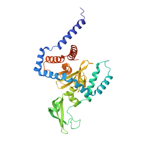
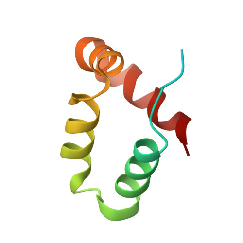
Structure of a peptide:N-glycanase-Rad23 complex: insight into the deglycosylation for denatured glycoproteins.

Lee, J.H.Choi, J.M.Lee, C.Yi, K.J.Cho, Y.

(2005) Proc Natl Acad Sci U S A 102: 9144-9149

  • DOI: https://doi.org/10.1073/pnas.0502082102
  • Primary Citation Related Structures: 
    1X3W, 1X3Z

  • PubMed Abstract: 

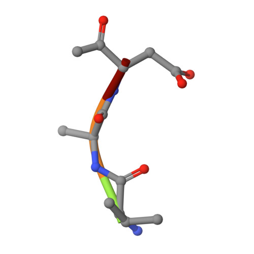
    In eukaryotes, misfolded proteins must be distinguished from correctly folded proteins during folding and transport processes by quality control systems. Yeast peptide:N-glycanase (yPNGase) specifically deglycosylates the denatured form of N-linked glycoproteins in the cytoplasm and assists proteasome-mediated glycoprotein degradation by forming a complex with 26S proteasome through DNA repair protein, yRad23. Here, we describe the crystal structures of a yPNGase and XPC-binding domain of yRad23 (yRad23XBD, residues 238-309) complex and of a yPNGase-yRad23XBD complex bound to a caspase inhibitor, Z-VAD-fmk. yPNGase is formed with three domains, a core domain containing a Cys-His-Asp triad, a Zn-binding domain, and a Rad23-binding domain. Both N- and C-terminal helices of yPNGase interact with yRad23 through extensive hydrophobic interactions. The active site of yPNGase is located in a deep cleft that is formed with residues conserved in all PNGase members, and three sugar molecules are bound to this cleft. Complex structures in conjunction with mutational analyses revealed that the walls of the cleft block access to the active site of yPNGase by native glycoprotein, whereas the cleft is sufficiently wide to accommodate denatured glycoprotein, thus explaining the specificity of PNGase for denatured substrates.


  • Organizational Affiliation
    • National Creative Research Center for Structural Biology and Department of Life Science, Pohang University of Science and Technology, Hyo-ja dong, San 31, Pohang, KyungBook, South Korea.

Macromolecule Content 

  • Total Structure Weight: 48.09 kDa 
  • Atom Count: 3,197 
  • Modeled Residue Count: 381 
  • Deposited Residue Count: 412 
  • Unique protein chains: 3

Macromolecules

Find similar proteins by:|  3D Structure
Entity ID: 1
MoleculeChains  Sequence LengthOrganismDetailsImage
peptide: N-glycanase335Saccharomyces cerevisiaeMutation(s): 0 
EC: 3.5.1.52
UniProt
Find proteins for Q02890 (Saccharomyces cerevisiae (strain ATCC 204508 / S288c))
Explore Q02890 
Go to UniProtKB:  Q02890
Entity Groups
Sequence Clusters30% Identity50% Identity70% Identity90% Identity95% Identity100% Identity
UniProt GroupQ02890
Sequence Annotations
Expand
Reference Sequence
Find similar proteins by:|  3D Structure
Entity ID: 2
MoleculeChains  Sequence LengthOrganismDetailsImage
UV excision repair protein RAD2372Saccharomyces cerevisiaeMutation(s): 0 
UniProt
Find proteins for P32628 (Saccharomyces cerevisiae (strain ATCC 204508 / S288c))
Explore P32628 
Go to UniProtKB:  P32628
Entity Groups
Sequence Clusters30% Identity50% Identity70% Identity90% Identity95% Identity100% Identity
UniProt GroupP32628
Sequence Annotations
Expand
Reference Sequence
Find similar proteins by:  Sequence   |   3D Structure  
Entity ID: 3
MoleculeChains  Sequence LengthOrganismDetailsImage
peptide PHQ-Val-Ala-Asp-CF0C [auth I]5N/AMutation(s): 0 

Oligosaccharides

Help  
Entity ID: 4
MoleculeChains Length2D Diagram GlycosylationD Interactions
beta-D-fructofuranose-(2-1)-alpha-D-glucopyranoseD [auth C],
E [auth D]
2N/A
Glycosylation Resources
GlyTouCan: G05551OP
GlyCosmos: G05551OP

Small Molecules

Ligands 1 Unique
IDChains Name / Formula / InChI Key2D Diagram3D Interactions
ZN

Query on ZN


Download:Ideal Coordinates CCD File
F [auth A]ZINC ION
Zn
PTFCDOFLOPIGGS-UHFFFAOYSA-N

Experimental Data & Validation

Experimental Data

  • Method: X-RAY DIFFRACTION
  • Resolution: 2.80 Å
  • R-Value Free:  0.270 (Depositor), 0.233 (DCC) 
  • R-Value Work:  0.236 (Depositor), 0.238 (DCC) 
  • R-Value Observed: 0.237 (Depositor) 
Space Group: P 31 2 1
Unit Cell:
Length ( Å )Angle ( ˚ )
a = 128.801α = 90
b = 128.801β = 90
c = 128.376γ = 120
Software Package:
Software NamePurpose
REFMACrefinement
HKL-2000data reduction
SCALEPACKdata scaling
CNSphasing

Structure Validation

View Full Validation Report


Entry History 

Deposition Data

Revision History  (Full details and data files)

  • Version 1.0: 2005-06-14
    Type: Initial release
  • Version 1.1: 2008-04-30
    Changes: Version format compliance
  • Version 1.2: 2011-07-13
    Changes: Atomic model, Database references, Derived calculations, Non-polymer description, Structure summary, Version format compliance
  • Version 1.3: 2012-12-12
    Changes: Other
  • Version 2.0: 2020-07-29
    Type: Remediation
    Reason: Carbohydrate remediation
    Changes: Atomic model, Data collection, Derived calculations, Non-polymer description, Structure summary
  • Version 2.1: 2024-10-09
    Changes: Data collection, Database references, Structure summary