1WDY | pdb_00001wdy

Crystal structure of ribonuclease


Experimental Data Snapshot

  • Method: X-RAY DIFFRACTION
  • Resolution: 1.80 Å
  • R-Value Free: 
    0.230 (Depositor) 
  • R-Value Work: 
    0.202 (Depositor) 
  • R-Value Observed: 
    0.203 (Depositor) 

wwPDB Validation 3D Report Full Report

Validation slider image for 1WDY

This is version 1.4 of the entry. See complete history

Literature

Structural basis for recognition of 2',5'-linked oligoadenylates by human ribonuclease L

Tanaka, N.Nakanishi, M.Kusakabe, Y.Goto, Y.Kitade, Y.Nakamura, K.T.

(2004) EMBO J 23: 3929-3938

  • DOI: https://doi.org/10.1038/sj.emboj.7600420
  • Primary Citation Related Structures: 
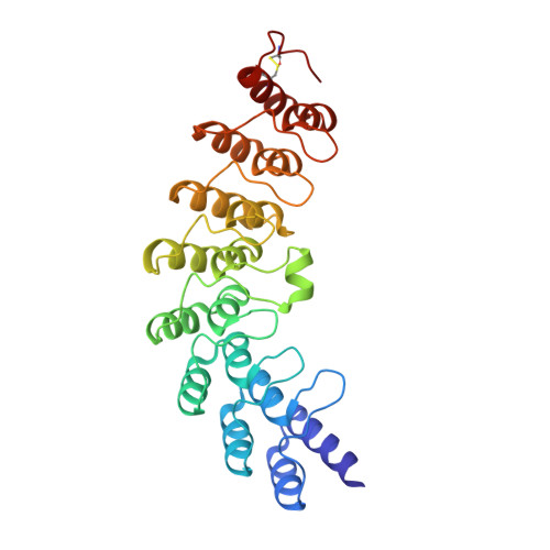
    1WDY

  • PubMed Abstract: 

    An interferon-induced endoribonuclease, ribonuclease L (RNase L), is implicated in both the molecular mechanism of action of interferon and the fundamental control of RNA stability in mammalian cells. RNase L is catalytically active only after binding to an unusual activator molecule containing a 5'-phosphorylated 2',5'-linked oligoadenylate (2-5A), in the N-terminal half. Here, we report the crystal structure of the N-terminal ankyrin repeat domain (ANK) of human RNase L complexed with the activator 2-5A. This is the first structural view of an ankyrin repeat structure directly interacting with a nucleic acid, rather than with a protein. The ANK domain folds into eight ankyrin repeat elements and forms an extended curved structure with a concave surface. The 2-5A molecule is accommodated at a concave site and directly interacts with ankyrin repeats 2-4. Interestingly, two structurally equivalent 2-5A binding motifs are found at repeats 2 and 4. The structural basis for 2-5A recognition by ANK is essential for designing stable 2-5As with a high likelihood of activating RNase L.


  • Organizational Affiliation
    • School of Pharmaceutical Sciences, Showa University, Hatanodai, Shinagawa-ku, Tokyo, Japan. ntanaka@pharm.showa-u.ac.jp

Macromolecule Content 

  • Total Structure Weight: 32.18 kDa 
  • Atom Count: 2,472 
  • Modeled Residue Count: 285 
  • Deposited Residue Count: 285 
  • Unique protein chains: 1

Macromolecules

Find similar proteins by:|  3D Structure
Entity ID: 1
MoleculeChains  Sequence LengthOrganismDetailsImage
2-5A-dependent ribonuclease285Homo sapiensMutation(s): 1 
EC: 3.1.26
UniProt & NIH Common Fund Data Resources
Find proteins for Q05823 (Homo sapiens)
Explore Q05823 
Go to UniProtKB:  Q05823
PHAROS:  Q05823
GTEx:  ENSG00000135828 
Entity Groups
Sequence Clusters30% Identity50% Identity70% Identity90% Identity95% Identity100% Identity
UniProt GroupQ05823
Sequence Annotations
Expand
Reference Sequence

Small Molecules

Ligands 1 Unique
IDChains Name / Formula / InChI Key2D Diagram3D Interactions
25A

Query on 25A


Download:Ideal Coordinates CCD File
B [auth A]5'-O-MONOPHOSPHORYLADENYLYL(2'->5')ADENYLYL(2'->5')ADENOSINE
C30 H38 N15 O19 P3
SIIZPVYVXNXXQG-UQTMIEBXSA-N

Experimental Data & Validation

Experimental Data

  • Method: X-RAY DIFFRACTION
  • Resolution: 1.80 Å
  • R-Value Free:  0.230 (Depositor) 
  • R-Value Work:  0.202 (Depositor) 
  • R-Value Observed: 0.203 (Depositor) 
Space Group: P 21 21 21
Unit Cell:
Length ( Å )Angle ( ˚ )
a = 63.203α = 90
b = 72.834β = 90
c = 82.634γ = 90
Software Package:
Software NamePurpose
REFMACrefinement
MOSFLMdata reduction
CCP4data scaling
AMoREphasing

Structure Validation

View Full Validation Report


Entry History 

Deposition Data

Revision History  (Full details and data files)

  • Version 1.0: 2004-10-05
    Type: Initial release
  • Version 1.1: 2008-04-30
    Changes: Version format compliance
  • Version 1.2: 2011-07-13
    Changes: Version format compliance
  • Version 1.3: 2021-11-10
    Changes: Database references, Derived calculations
  • Version 1.4: 2024-11-20
    Changes: Data collection, Structure summary