1UYU

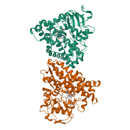
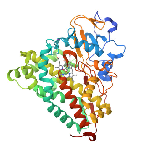
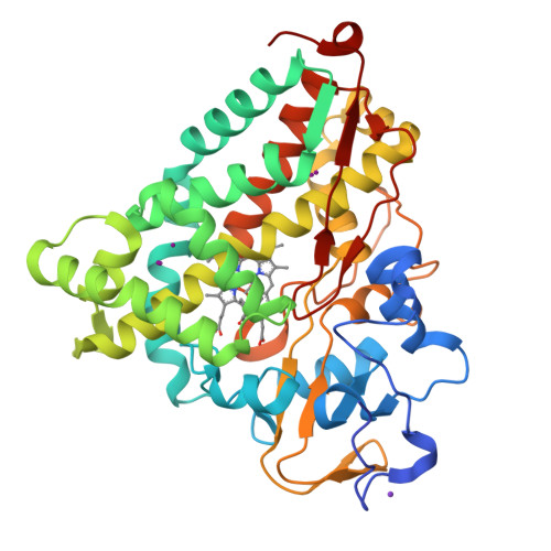
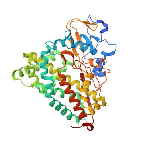
Xenon COMPLEX OF wildtype P450CAM FROM PSEUDOMONAS PUTIDA


Experimental Data Snapshot

  • Method: X-RAY DIFFRACTION
  • Resolution: 2.00 Å
  • R-Value Free: 0.236 
  • R-Value Work: 0.187 
  • R-Value Observed: 0.190 

Starting Model: experimental
View more details

wwPDB Validation   3D Report Full Report


Ligand Structure Quality Assessment 


This is version 1.3 of the entry. See complete history


Literature

A Survey of Active Site Access Channels in Cytochromes P450

Wade, R.C.Winn, P.J.Schlichting, I.

(2004) J Inorg Biochem 98: 1175

  • DOI: https://doi.org/10.1016/j.jinorgbio.2004.02.007
  • Primary Citation of Related Structures:  
    1UYU

  • PubMed Abstract: 

    In cytochrome P450s, the active site is situated deep inside the protein next to the heme cofactor, and is often completely isolated from the surrounding solvent. To identify routes by which substrates may enter into and products exit from the active site, random expulsion molecular dynamics simulations were performed for three cytochrome P450s: CYP101, CYP102A1 and CYP107A1 [J. Mol. Biol. 303 (2000) 797; Proc. Natl. Acad. Sci. USA 99 (2002) 5361]. Amongst the different pathways identified, one pathway was found to be common to all three cytochrome P450s although the mechanism of ligand passage along it was different in each case and apparently adapted to the substrate specificity of the enzyme. Recently, a number of new crystal structures of cytochrome P450s have been solved. Here, we analyse the open channels leading to the active site that these structures reveal. We find that in addition to showing the common pathway, they provide experimental evidence for the existence of three additional channels that were identified by simulation. We also discuss how the location of xenon binding sites in CYP101 suggests a role for one of the pathways identified by molecular dynamics simulations as a route for gaseous species, such as oxygen, to access the active site.


  • Organizational Affiliation

    Molecular and Cellular Modeling, EML Research, Villa Bosch, Schloss-Wolfsbrunnenweg 33, 69118, Heidelberg, Germany. rebecca.wade@eml-r.villa-bosch.de


Macromolecules
Find similar proteins by:  (by identity cutoff)  |  3D Structure
Entity ID: 1
MoleculeChains Sequence LengthOrganismDetailsImage
CYTOCHROME P450-CAM
A, B
414Pseudomonas putidaMutation(s): 0 
EC: 1.14.15.1
UniProt
Find proteins for P00183 (Pseudomonas putida)
Explore P00183 
Go to UniProtKB:  P00183
Entity Groups  
Sequence Clusters30% Identity50% Identity70% Identity90% Identity95% Identity100% Identity
UniProt GroupP00183
Sequence Annotations
Expand
  • Reference Sequence
Small Molecules
Ligands 4 Unique
IDChains Name / Formula / InChI Key2D Diagram3D Interactions
HEM
Query on HEM

Download Ideal Coordinates CCD File 
C [auth A],
J [auth B]
PROTOPORPHYRIN IX CONTAINING FE
C34 H32 Fe N4 O4
KABFMIBPWCXCRK-RGGAHWMASA-L
CAM
Query on CAM

Download Ideal Coordinates CCD File 
D [auth A],
K [auth B]
CAMPHOR
C10 H16 O
DSSYKIVIOFKYAU-XCBNKYQSSA-N
XE
Query on XE

Download Ideal Coordinates CCD File 
F [auth A]
G [auth A]
H [auth A]
I [auth A]
N [auth B]
F [auth A],
G [auth A],
H [auth A],
I [auth A],
N [auth B],
O [auth B],
P [auth B],
Q [auth B]
XENON
Xe
FHNFHKCVQCLJFQ-UHFFFAOYSA-N
K
Query on K

Download Ideal Coordinates CCD File 
E [auth A],
L [auth B],
M [auth B]
POTASSIUM ION
K
NPYPAHLBTDXSSS-UHFFFAOYSA-N
Experimental Data & Validation

Experimental Data

  • Method: X-RAY DIFFRACTION
  • Resolution: 2.00 Å
  • R-Value Free: 0.236 
  • R-Value Work: 0.187 
  • R-Value Observed: 0.190 
  • Space Group: P 1 21 1
Unit Cell:
Length ( Å )Angle ( ˚ )
a = 67.2α = 90
b = 62.3β = 90.5
c = 95.3γ = 90
Software Package:
Software NamePurpose
REFMACrefinement
XDSdata reduction
XSCALEdata scaling

Structure Validation

View Full Validation Report


Ligand Structure Quality Assessment 


Entry History 

Deposition Data

Revision History  (Full details and data files)

  • Version 1.0: 2005-03-07
    Type: Initial release
  • Version 1.1: 2012-08-22
    Changes: Database references, Other, Structure summary, Version format compliance
  • Version 1.2: 2019-04-03
    Changes: Data collection, Experimental preparation, Other
  • Version 1.3: 2023-12-13
    Changes: Data collection, Database references, Derived calculations, Other, Refinement description