1UCB | pdb_00001ucb

STRUCTURE OF UNCOMPLEXED FAB COMPARED TO COMPLEX (1CLY, 1CLZ)


Experimental Data Snapshot

  • Method: X-RAY DIFFRACTION
  • Resolution: 2.50 Å
  • R-Value Free: 
    0.276 (Depositor) 
  • R-Value Work: 
    0.200 (Depositor), 0.183 (DCC) 
  • R-Value Observed: 
    0.200 (Depositor) 

Starting Models: experimental
View more details

wwPDB Validation 3D Report Full Report

Validation slider image for 1UCB

This is version 1.4 of the entry. See complete history

Literature

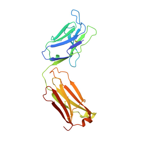
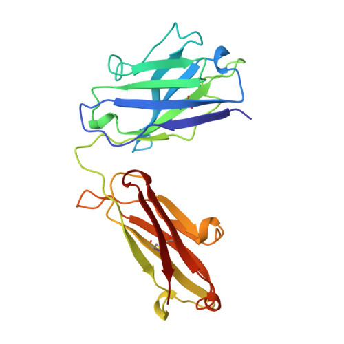
X-ray structure of the uncomplexed anti-tumor antibody BR96 and comparison with its antigen-bound form.

Sheriff, S.Chang, C.Y.Jeffrey, P.D.Bajorath, J.

(1996) J Mol Biology 259: 938-946

  • DOI: https://doi.org/10.1006/jmbi.1996.0371
  • Primary Citation Related Structures: 
    1UCB

  • PubMed Abstract: 

    The X-ray structure of the uncomplexed human chimeric Fab' of the anti-tumor antibody BR96 has been determined at 2.6 A resolution. The structure has been compared with Lewis Y antigen-complexed structures of BR96 which were determined previously. The comparison reveals segmental motions and/or conformational rearrangements of three CDR loops (L1, L3, and H2), whereas CDR H3 does not undergo changes upon complexation despite its significant main-chain contacts to the carbohydrate antigen. In light of the uncomplexed chimeric Fab' structure reported here, the previously observed high mobility of the CL:CH1 domains of the complexed chimeric BR96 Fab is rationalized as a "swinging" motion approximately about the axis of the elbow bend.


  • Organizational Affiliation
    • Bristol-Myers Squibb Pharmaceutical Research Institute, Princeton, NJ 08543-4000, USA.

Macromolecule Content 

  • Total Structure Weight: 47.78 kDa 
  • Atom Count: 3,378 
  • Modeled Residue Count: 435 
  • Deposited Residue Count: 438 
  • Unique protein chains: 2

Macromolecules

Find similar proteins by:|  3D Structure
Entity ID: 1
MoleculeChains  Sequence LengthOrganismDetailsImage
CHIMERIC HUMAN/MOUSE IGG FAB FRAGMENT BR96A [auth L]219Homo sapiensMutation(s): 0 
UniProt & NIH Common Fund Data Resources
Find proteins for P01834 (Homo sapiens)
Explore P01834 
Go to UniProtKB:  P01834
PHAROS:  P01834
Entity Groups
Sequence Clusters30% Identity50% Identity70% Identity90% Identity95% Identity100% Identity
UniProt GroupP01834
Sequence Annotations
Expand
Reference Sequence
Find similar proteins by:|  3D Structure
Entity ID: 2
MoleculeChains  Sequence LengthOrganismDetailsImage
CHIMERIC HUMAN/MOUSE IGG FAB FRAGMENT BR96B [auth H]219Homo sapiensMutation(s): 0 

Small Molecules

Ligands 1 Unique
IDChains Name / Formula / InChI Key2D Diagram3D Interactions
SO4

Query on SO4


Download:Ideal Coordinates CCD File
C [auth L]SULFATE ION
O4 S
QAOWNCQODCNURD-UHFFFAOYSA-L

Experimental Data & Validation

Experimental Data

  • Method: X-RAY DIFFRACTION
  • Resolution: 2.50 Å
  • R-Value Free:  0.276 (Depositor) 
  • R-Value Work:  0.200 (Depositor), 0.183 (DCC) 
  • R-Value Observed: 0.200 (Depositor) 
Space Group: P 21 21 2
Unit Cell:
Length ( Å )Angle ( ˚ )
a = 61.1α = 90
b = 174.3β = 90
c = 45.6γ = 90
Software Package:
Software NamePurpose
XDSdata scaling
XSCALEdata scaling
BRUTEmodel building
MERLOTphasing
X-PLORrefinement
XDSdata reduction
BRUTEphasing

Structure Validation

View Full Validation Report


Entry History 

Deposition Data

Revision History  (Full details and data files)

  • Version 1.0: 1997-03-12
    Type: Initial release
  • Version 1.1: 2008-03-24
    Changes: Version format compliance
  • Version 1.2: 2011-07-13
    Changes: Version format compliance
  • Version 1.3: 2023-08-09
    Changes: Database references, Derived calculations, Refinement description
  • Version 1.4: 2024-11-20
    Changes: Data collection, Structure summary